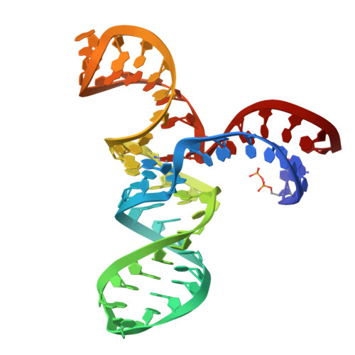
9FN3 | pdb_00009fn3

Crystal structure of the alkyltransferase ribozyme SAMURI co-crystalized with ProSeDMA


Experimental Data Snapshot

  • Method: X-RAY DIFFRACTION
  • Resolution: 2.90 Å
  • R-Value Free: 
    0.224 (Depositor), 0.243 (DCC) 
  • R-Value Work: 
    0.201 (Depositor), 0.214 (DCC) 
  • R-Value Observed: 
    0.202 (Depositor) 

wwPDB Validation   3D Report Full Report


Ligand Structure Quality Assessment 


This is version 2.0 of the entry. See complete history


Literature

Structure and catalytic activity of the SAM-utilizing ribozyme SAMURI.

Chen, H.A.Okuda, T.Lenz, A.K.Scheitl, C.P.M.Schindelin, H.Hobartner, C.

(2025) Nat Chem Biol 

  • DOI: https://doi.org/10.1038/s41589-024-01808-w
  • Primary Citation of Related Structures:  
    9FN2, 9FN3

  • PubMed Abstract: 

    Ribozymes that catalyze site-specific RNA modification have recently gained increasing interest for their ability to mimic methyltransferase enzymes and for their application to install molecular tags. Recently, we reported SAMURI as a site-specific alkyltransferase ribozyme using S-adenosylmethionine (SAM) or a stabilized analog to transfer a methyl or propargyl group to N 3 of an adenosine. Here, we report the crystal structures of SAMURI in the postcatalytic state. The structures reveal a three-helix junction with the catalytic core folded into four stacked layers, harboring the cofactor and the modified nucleotide. Detailed structure-activity analyses explain the cofactor scope and the structural basis for site selectivity. A structural comparison of SAMURI with SAM riboswitches sheds light on how the synthetic ribozyme overcomes the strategies of natural riboswitches to avoid self-methylation. Our results suggest that SAM and its analogs may serve as substrates for various RNA-catalyzed reactions, for which the corresponding ribozymes remain to be identified.


  • Organizational Affiliation

    Institute of Organic Chemistry, Julius-Maximilians-Universität Würzburg, Würzburg, Germany.


Macromolecules
Find similar nucleic acids by:  (by identity cutoff)  |  3D Structure
Entity ID: 1
MoleculeChains LengthOrganismImage
SAMURI-ProSeDMA
A, B
58synthetic construct
Sequence Annotations
Expand
  • Reference Sequence
Small Molecules
Experimental Data & Validation

Experimental Data

  • Method: X-RAY DIFFRACTION
  • Resolution: 2.90 Å
  • R-Value Free:  0.224 (Depositor), 0.243 (DCC) 
  • R-Value Work:  0.201 (Depositor), 0.214 (DCC) 
  • R-Value Observed: 0.202 (Depositor) 
Space Group: P 42
Unit Cell:
Length ( Å )Angle ( ˚ )
a = 75.444α = 90
b = 75.444β = 90
c = 72.251γ = 90
Software Package:
Software NamePurpose
PHENIXrefinement
XDSdata scaling
XDSdata reduction
AutoSolphasing
Cootmodel building

Structure Validation

View Full Validation Report


Ligand Structure Quality Assessment 


Entry History & Funding Information

Deposition Data


Funding OrganizationLocationGrant Number
German Research Foundation (DFG)Germany--
European Research Council (ERC)European Union--

Revision History  (Full details and data files)

  • Version 1.0: 2025-02-05
    Type: Initial release
  • Version 2.0: 2025-02-12
    Type: Coordinate replacement
    Reason: Ligand geometry
    Changes: Advisory, Atomic model, Author supporting evidence, Data collection, Database references, Derived calculations, Refinement description, Structure summary