9FH8 | pdb_00009fh8

Crystal structure of the SPD-2 domain of Apis dorsata CEP192


Experimental Data Snapshot

  • Method: X-RAY DIFFRACTION
  • Resolution: 3.50 Å
  • R-Value Free: 
    0.313 (Depositor), 0.313 (DCC) 
  • R-Value Work: 
    0.287 (Depositor), 0.287 (DCC) 
  • R-Value Observed: 
    0.289 (Depositor) 

Starting Model: in silico
View more details

wwPDB Validation   3D Report Full Report


This is version 1.1 of the entry. See complete history


Literature

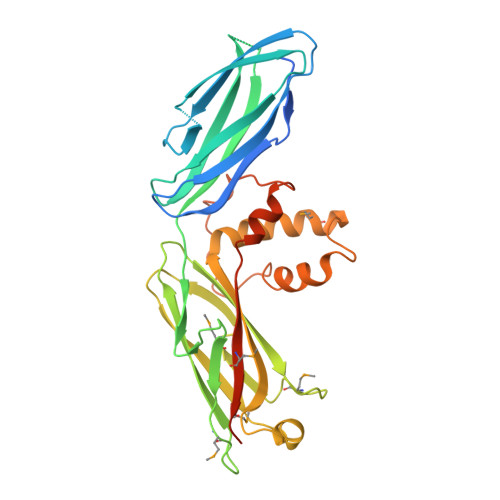
The conserved Spd-2/CEP192 domain adopts a unique protein fold to promote centrosome scaffold assembly.

Hu, L.Wainman, A.Andreeva, A.Apizi, M.Alvarez-Rodrigo, I.Wong, S.S.Saurya, S.Sheppard, D.Cottee, M.Johnson, S.Lea, S.M.Raff, J.W.van Breugel, M.Feng, Z.

(2025) Sci Adv 11: eadr5744-eadr5744

  • DOI: https://doi.org/10.1126/sciadv.adr5744
  • Primary Citation of Related Structures:  
    9C72, 9FH8, 9FU8

  • PubMed Abstract: 

    Centrosomes form when centrioles assemble pericentriolar material (PCM) around themselves. Spd-2/CEP192 proteins, defined by a conserved "Spd-2 domain" (SP2D) comprising two closely spaced AspM-Spd-2-Hydin (ASH) domains, play a critical role in centrosome assembly. Here, we show that the SP2D does not target Drosophila Spd-2 to centrosomes but rather promotes PCM scaffold assembly. Crystal structures of the human and honeybee SP2D reveal an unusual "extended cradle" structure mediated by a conserved interaction interface between the two ASH domains. Mutations predicted to perturb this interface, including a human mutation associated with male infertility and Mosaic Variegated Aneuploidy, disrupt PCM scaffold assembly in flies. The SP2D is monomeric in solution, but the Drosophila SP2D can form higher-order oligomers upon phosphorylation by PLK1 (Polo-like kinase 1). Crystal-packing interactions and AlphaFold predictions suggest how SP2Ds might self-assemble, and mutations associated with one such potential dimerization interface markedly perturb SP2D oligomerization in vitro and PCM scaffold assembly in vivo.


  • Organizational Affiliation
    • State Key Laboratory of Genetic Engineering, School of Life Sciences, Fudan University, Shanghai, China.

Macromolecules
Find similar proteins by:  (by identity cutoff)  |  3D Structure
Entity ID: 1
MoleculeChains Sequence LengthOrganismDetailsImage
Centrosomal protein 192 (CEP192)
A, B, C, D, E
A, B, C, D, E, F, G, H
341Apis dorsataMutation(s): 0 
EC: 3.1.1.4
UniProt
Find proteins for A0A7M7MR83 (Apis mellifera)
Explore A0A7M7MR83 
Go to UniProtKB:  A0A7M7MR83
Entity Groups  
Sequence Clusters30% Identity50% Identity70% Identity90% Identity95% Identity100% Identity
UniProt GroupA0A7M7MR83
Sequence Annotations
Expand
  • Reference Sequence
Small Molecules
Modified Residues  1 Unique
IDChains TypeFormula2D DiagramParent
MSE
Query on MSE
A, B, C, D, E
A, B, C, D, E, F, G, H
L-PEPTIDE LINKINGC5 H11 N O2 SeMET
Experimental Data & Validation

Experimental Data

  • Method: X-RAY DIFFRACTION
  • Resolution: 3.50 Å
  • R-Value Free:  0.313 (Depositor), 0.313 (DCC) 
  • R-Value Work:  0.287 (Depositor), 0.287 (DCC) 
  • R-Value Observed: 0.289 (Depositor) 
Space Group: I 41
Unit Cell:
Length ( Å )Angle ( ˚ )
a = 183.3α = 90
b = 183.3β = 90
c = 190.044γ = 90
Software Package:
Software NamePurpose
PHENIXrefinement
XDSdata reduction
Aimlessdata scaling
PHASERphasing

Structure Validation

View Full Validation Report


Entry History & Funding Information

Deposition Data


Funding OrganizationLocationGrant Number
Medical Research Council (MRC, United Kingdom)United KingdomMC_UP_1201/3

Revision History  (Full details and data files)

  • Version 1.0: 2025-03-26
    Type: Initial release
  • Version 1.1: 2025-04-02
    Changes: Database references