9F80 | pdb_00009f80

Crystal structure of Rv2242 regulator C-terminal fragment (161-414)


Experimental Data Snapshot

  • Method: X-RAY DIFFRACTION
  • Resolution: 2.03 Å
  • R-Value Free: 
    0.254 (Depositor), 0.253 (DCC) 
  • R-Value Work: 
    0.196 (Depositor), 0.206 (DCC) 

wwPDB Validation 3D Report Full Report

Validation slider image for 9F80

This is version 1.0 of the entry. See complete history

Literature

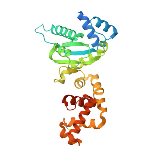
Domain architecture of the Mycobacterium tuberculosis MabR ( Rv2242 ), a member of the PucR transcription factor family.

Megalizzi, V.Tanina, A.Grosse, C.Mirgaux, M.Legrand, P.Dias Mirandela, G.Wohlkonig, A.Bifani, P.Wintjens, R.

(2024) Heliyon 10: e40494-e40494

  • DOI: https://doi.org/10.1016/j.heliyon.2024.e40494
  • Primary Citation Related Structures: 
    9F80, 9FB1

  • PubMed Abstract: 

    MabR ( Rv2242 ), a PucR-type transcription factor, plays a crucial role in regulating mycolic acid biosynthesis in Mycobacterium tuberculosis . To understand its regulatory mechanisms, we determined the crystal structures of its N-terminal and C-terminal domains. The N-terminal domain adopts a globin-like fold, while the C-terminal domain comprises an α/β GGDEF domain and an all-α effector domain with a helix-turn-helix DNA-binding motif. This unique domain combination is specific to Actinomycetes . Biochemical and computational studies suggest that full-length MabR forms both dimeric and tetrameric assemblies in solution. Structural analysis revealed two distinct dimerization interfaces within the N- and C-terminal domains, further supporting a tetrameric organization. These findings provide valuable insights into the domain architecture, oligomeric state, and potential regulatory mechanisms of MabR.


  • Organizational Affiliation
    • Unit of Microbiology, Bioorganic and Macromolecular Chemistry, Department of Research in Drug Development, Faculty of Pharmacy, Université Libre de Bruxelles, Belgium.

Macromolecule Content 

  • Total Structure Weight: 29.25 kDa 
  • Atom Count: 1,834 
  • Modeled Residue Count: 243 
  • Deposited Residue Count: 275 
  • Unique protein chains: 1

Macromolecules

Find similar proteins by:|  3D Structure
Entity ID: 1
MoleculeChains  Sequence LengthOrganismDetailsImage
Uncharacterized protein Rv2242275Mycobacterium tuberculosis H37RvMutation(s): 0 
Gene Names: Rv2242MTCY427.23
UniProt
Find proteins for P9WPH5 (Mycobacterium tuberculosis (strain ATCC 25618 / H37Rv))
Explore P9WPH5 
Go to UniProtKB:  P9WPH5
Entity Groups
Sequence Clusters30% Identity50% Identity70% Identity90% Identity95% Identity100% Identity
UniProt GroupP9WPH5
Sequence Annotations
Expand
Reference Sequence

Small Molecules

Experimental Data & Validation

Experimental Data

  • Method: X-RAY DIFFRACTION
  • Resolution: 2.03 Å
  • R-Value Free:  0.254 (Depositor), 0.253 (DCC) 
  • R-Value Work:  0.196 (Depositor), 0.206 (DCC) 
Space Group: C 2 2 21
Unit Cell:
Length ( Å )Angle ( ˚ )
a = 63.04α = 90
b = 75.28β = 90
c = 139.52γ = 90
Software Package:
Software NamePurpose
REFMACrefinement
XDSdata reduction
XDSdata scaling
SHELXDphasing
PHASERphasing
MOLREPphasing

Structure Validation

View Full Validation Report


Entry History 

& Funding Information

Deposition Data


Funding OrganizationLocationGrant Number
Fonds National de la Recherche Scientifique (FNRS)BelgiumCR40003580

Revision History  (Full details and data files)

  • Version 1.0: 2024-12-18
    Type: Initial release