9F6T | pdb_00009f6t

cryoEM structure of Asgard tubulin heterodimer AtubA/B with GTP


Experimental Data Snapshot

  • Method: ELECTRON MICROSCOPY
  • Resolution: 3.50 Å
  • Aggregation State: PARTICLE 
  • Reconstruction Method: SINGLE PARTICLE 

wwPDB Validation 3D Report Full Report

Validation slider image for 9F6T

This is version 1.1 of the entry. See complete history

Literature

Microtubules in Asgard archaea.

Wollweber, F.Xu, J.Ponce-Toledo, R.I.Marxer, F.Rodrigues-Oliveira, T.Possnecker, A.Luo, Z.H.Malit, J.J.L.Kokhanovska, A.Wieczorek, M.Schleper, C.Pilhofer, M.

(2025) Cell 188: 2451

  • DOI: https://doi.org/10.1016/j.cell.2025.02.027
  • Primary Citation Related Structures: 
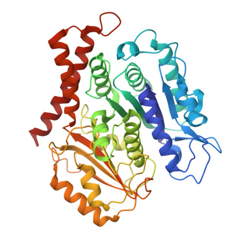
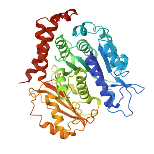
    9F6T, 9F6U, 9F6V, 9HXC

  • PubMed Abstract: 

    Microtubules are a hallmark of eukaryotes. Archaeal and bacterial homologs of tubulins typically form homopolymers and non-tubular superstructures. The origin of heterodimeric tubulins assembling into microtubules remains unclear. Here, we report the discovery of microtubule-forming tubulins in Asgard archaea, the closest known relatives of eukaryotes. These Asgard tubulins (AtubA/B) are closely related to eukaryotic α/β-tubulins and the enigmatic bacterial tubulins BtubA/B. Proteomics of Candidatus Lokiarchaeum ossiferum showed that AtubA/B were highly expressed. Cryoelectron microscopy structures demonstrate that AtubA/B form eukaryote-like heterodimers, which assembled into 5-protofilament bona fide microtubules in vitro. The additional paralog AtubB2 lacks a nucleotide-binding site and competitively displaced AtubB. These AtubA/B2 heterodimers polymerized into 7-protofilament non-canonical microtubules. In a sub-population of Ca. Lokiarchaeum ossiferum cells, cryo-tomography revealed tubular structures, while expansion microscopy identified AtubA/B cytoskeletal assemblies. Our findings suggest a pre-eukaryotic origin of microtubules and provide a framework for understanding the fundamental principles of microtubule assembly.


  • Organizational Affiliation
    • Department of Biology, Institute of Molecular Biology & Biophysics, Eidgenössische Technische Hochschule Zürich, Otto-Stern-Weg 5, 8093 Zürich, Switzerland.

Macromolecule Content 

  • Total Structure Weight: 93.97 kDa 
  • Atom Count: 6,574 
  • Modeled Residue Count: 845 
  • Deposited Residue Count: 847 
  • Unique protein chains: 2

Macromolecules

Find similar proteins by:|  3D Structure
Entity ID: 1
MoleculeChains  Sequence LengthOrganismDetailsImage
Asgard tubulin A (AtubA) from Candidatus Lokiarchaeum ossiferum423Candidatus Lokiarchaeum ossiferumMutation(s): 0 
UniProt
Find proteins for A0A7W7YIU5 (Prosthecobacter dejongeii)
Explore A0A7W7YIU5 
Go to UniProtKB:  A0A7W7YIU5
Entity Groups
Sequence Clusters30% Identity50% Identity70% Identity90% Identity95% Identity100% Identity
UniProt GroupA0A7W7YIU5
Sequence Annotations
Expand
Reference Sequence
Find similar proteins by:|  3D Structure
Entity ID: 2
MoleculeChains  Sequence LengthOrganismDetailsImage
Asgard tubulin B (AtubB) from Candidatus Lokiarchaeum ossiferum424Candidatus Lokiarchaeum ossiferumMutation(s): 0 
UniProt
Find proteins for A0A7W7YJ10 (Prosthecobacter dejongeii)
Explore A0A7W7YJ10 
Go to UniProtKB:  A0A7W7YJ10
Entity Groups
Sequence Clusters30% Identity50% Identity70% Identity90% Identity95% Identity100% Identity
UniProt GroupA0A7W7YJ10
Sequence Annotations
Expand
Reference Sequence

Experimental Data & Validation

Experimental Data

  • Method: ELECTRON MICROSCOPY
  • Resolution: 3.50 Å
  • Aggregation State: PARTICLE 
  • Reconstruction Method: SINGLE PARTICLE 

Structure Validation

View Full Validation Report


Entry History 

& Funding Information

Deposition Data


Funding OrganizationLocationGrant Number
European Research Council (ERC)European Union101000232

Revision History  (Full details and data files)

  • Version 1.0: 2025-04-02
    Type: Initial release
  • Version 1.1: 2025-05-14
    Changes: Data collection, Database references