9F23 | pdb_00009f23

DARPin eGFP complex DP2 (2G156)


Experimental Data Snapshot

  • Method: X-RAY DIFFRACTION
  • Resolution: 1.59 Å
  • R-Value Free: 
    0.212 (Depositor), 0.217 (DCC) 
  • R-Value Work: 
    0.174 (Depositor), 0.185 (DCC) 
  • R-Value Observed: 
    0.176 (Depositor) 

Starting Model: experimental
View more details

wwPDB Validation   3D Report Full Report


Ligand Structure Quality Assessment 


This is version 1.1 of the entry. See complete history


Literature

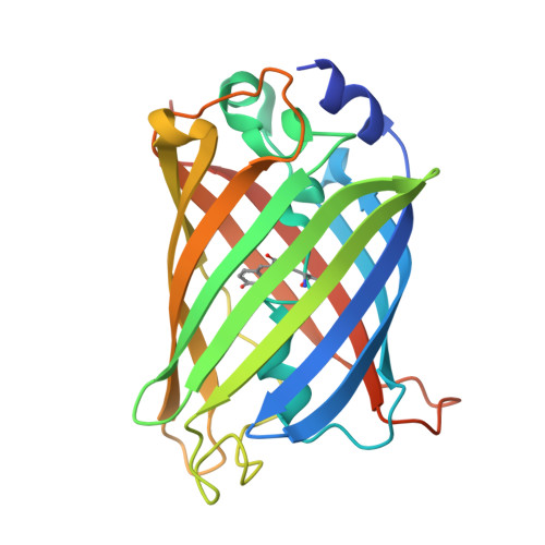
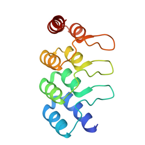
Molecular features defining the efficiency of bioPROTACs.

Winkelvoss, D.Vukovic, D.Hanny, A.C.Riermeier, L.Udovcic, A.Kolibius, J.Honegger, A.Mittl, P.R.E.Michel, E.Hansen, S.Pluckthun, A.

(2025) Commun Biol 8: 946-946

  • DOI: https://doi.org/10.1038/s42003-025-08352-w
  • Primary Citation of Related Structures:  
    9F22, 9F23, 9F24

  • PubMed Abstract: 

    BioPROTACs consist of a target-binding unit and a component of the ubiquitin-proteasome system. However, the specific biophysical features influencing their effectiveness are poorly understood. We investigated the design principles defining the target-binding moiety of bioPROTACs. We used our recently developed assay for accurately measuring degradation rates, based on microinjection and live-cell microscopy, independent of other confounding factors like biosynthesis and transport. We used a very efficient UPS interaction domain from CHIP E3 ligase, and 9 different well-characterized DARPins to test degradation of the proof-of-principle target eGFP. All but two DARPins work well in this context, one sterically preventing E2 binding in the complex, the other overlapping with the target ubiquitination epitope. Affinity and thermodynamic stability of the binders had only a modest role. BioPROTACs constructed in this way were able to degrade eGFP catalytically. DARPins by themselves could also accelerate degradation of bound GFP, using other cellular E3 systems, but in a non-catalytic manner. The most important factor for efficient degradation by a bioPROTAC in trans is the correct orientation of the complex for target ubiquitination and presentation to the proteasome, still to be determined empirically. The strategies developed here show an efficient pathway to characterize and optimize such systems.


  • Organizational Affiliation
    • Department of Biochemistry, University of Zurich, Zurich, Switzerland.

Macromolecules
Find similar proteins by:  (by identity cutoff)  |  3D Structure
Entity ID: 1
MoleculeChains Sequence LengthOrganismDetailsImage
Green fluorescent protein
A, C
243Aequorea victoriaMutation(s): 3 
Gene Names: GFP
UniProt
Find proteins for P42212 (Aequorea victoria)
Explore P42212 
Go to UniProtKB:  P42212
Entity Groups  
Sequence Clusters30% Identity50% Identity70% Identity90% Identity95% Identity100% Identity
UniProt GroupP42212
Sequence Annotations
Expand
  • Reference Sequence
Find similar proteins by:  (by identity cutoff)  |  3D Structure
Entity ID: 2
MoleculeChains Sequence LengthOrganismDetailsImage
DARPin DP2
B, D
128synthetic constructMutation(s): 0 
Entity Groups  
Sequence Clusters30% Identity50% Identity70% Identity90% Identity95% Identity100% Identity
Sequence Annotations
Expand
  • Reference Sequence
Small Molecules
Ligands 5 Unique
IDChains Name / Formula / InChI Key2D Diagram3D Interactions
PXN
Query on PXN

Download Ideal Coordinates CCD File 
R [auth C](2S)-1-[3-{[(2R)-2-hydroxypropyl]oxy}-2,2-bis({[(2R)-2-hydroxypropyl]oxy}methyl)propoxy]propan-2-ol
C17 H36 O8
GXEZGLLPFFKHGE-FPCVCCKLSA-N
A1H87
Query on A1H87

Download Ideal Coordinates CCD File 
E [auth A],
Q [auth C]
2-[[(2S)-2-oxidanylpropoxy]methyl]-2-[[(2S)-2-[(2S)-2-oxidanylpropoxy]propoxy]methyl]propane-1,3-diol
C14 H30 O7
GYGFRXYZKNFZRH-AVGNSLFASA-N
TRS
Query on TRS

Download Ideal Coordinates CCD File 
BA [auth D]
CA [auth D]
DA [auth D]
N [auth B]
O [auth B]
BA [auth D],
CA [auth D],
DA [auth D],
N [auth B],
O [auth B],
P [auth B]
2-AMINO-2-HYDROXYMETHYL-PROPANE-1,3-DIOL
C4 H12 N O3
LENZDBCJOHFCAS-UHFFFAOYSA-O
EDO
Query on EDO

Download Ideal Coordinates CCD File 
AA [auth D]
F [auth A]
G [auth A]
H [auth A]
J [auth B]
AA [auth D],
F [auth A],
G [auth A],
H [auth A],
J [auth B],
K [auth B],
L [auth B],
M [auth B],
S [auth C],
T [auth C],
U [auth C],
V [auth C],
W [auth C],
Y [auth D],
Z [auth D]
1,2-ETHANEDIOL
C2 H6 O2
LYCAIKOWRPUZTN-UHFFFAOYSA-N
NA
Query on NA

Download Ideal Coordinates CCD File 
I [auth A],
X [auth C]
SODIUM ION
Na
FKNQFGJONOIPTF-UHFFFAOYSA-N
Modified Residues  1 Unique
IDChains TypeFormula2D DiagramParent
CRO
Query on CRO
A, C
L-PEPTIDE LINKINGC15 H17 N3 O5THR, TYR, GLY
Experimental Data & Validation

Experimental Data

  • Method: X-RAY DIFFRACTION
  • Resolution: 1.59 Å
  • R-Value Free:  0.212 (Depositor), 0.217 (DCC) 
  • R-Value Work:  0.174 (Depositor), 0.185 (DCC) 
  • R-Value Observed: 0.176 (Depositor) 
Space Group: P 1 21 1
Unit Cell:
Length ( Å )Angle ( ˚ )
a = 61.87α = 90
b = 95.16β = 115.76
c = 69.47γ = 90
Software Package:
Software NamePurpose
REFMACrefinement
XDSdata reduction
XSCALEdata scaling
MOLREPphasing

Structure Validation

View Full Validation Report


Ligand Structure Quality Assessment 


Entry History & Funding Information

Deposition Data


Funding OrganizationLocationGrant Number
Swiss National Science FoundationSwitzerland--

Revision History  (Full details and data files)

  • Version 1.0: 2024-10-23
    Type: Initial release
  • Version 1.1: 2025-11-12
    Changes: Database references