9E92 | pdb_00009e92

Acanthamoeba Polyphaga Mimivirus R699


Experimental Data Snapshot

  • Method: X-RAY DIFFRACTION
  • Resolution: 1.50 Å
  • R-Value Free: 
    0.189 (Depositor), 0.191 (DCC) 
  • R-Value Work: 
    0.166 (Depositor), 0.167 (DCC) 

Starting Model: experimental
View more details

wwPDB Validation 3D Report Full Report

Validation slider image for 9E92

Ligand Structure Quality Assessment 


This is version 1.1 of the entry. See complete history

Literature

Structural basis of collagen glucosyltransferase function and its serendipitous role in kojibiose synthesis.

Kim, J.S.Chen, Z.Espinosa Garcia, S.A.Buhlheller, C.Zhang, B.Richards, S.J.Chen, T.Wu, J.Bruntz, R.C.Gilliam, M.E.Yamauchi, M.Liang, B.Guo, H.

(2025) Nat Commun 16: 6704-6704

  • DOI: https://doi.org/10.1038/s41467-025-61973-x
  • Primary Citation Related Structures: 
    9DYT, 9DZS, 9E92

  • PubMed Abstract: 

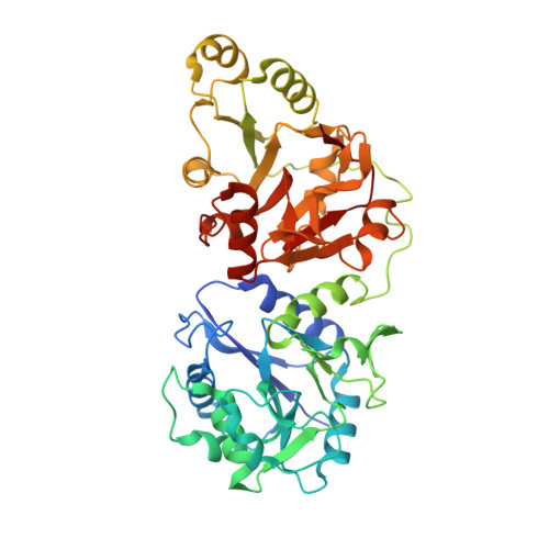
    Collagen glucosyltransferases catalyze collagen glucosylation critical for biology and diseases, yet their structural regulation remains unclear. Here, we report crystal structures of a mimiviral collagen glucosyltransferase in its apo form and in complexes with uridine diphosphate (UDP) and the disaccharide product. We reveal that the enzyme forms a homodimer, stabilized by a loop from one subunit locking into a cleft on the other, enabling UDP-glucose binding cooperativity and enzymatic activity, a property conserved in the human homolog. The structures support an induced fit model for UDP interaction. The dimerization also forms an extended cleft flanked by two active sites, likely facilitating collagen recognition. Unexpectedly, the mimiviral enzyme also synthesizes a prebiotic disaccharide kojibiose. An elongated pocket near the active site allows the enzyme to use UDP-glucose and glucose for kojibiose production. We confirm the enzyme's kojibiose synthesis activity in vitro and in vivo. These insights inform glucosyltransferase function and open new avenues for inhibitor development and kojibiose biosynthesis.


  • Organizational Affiliation
    • Department of Molecular and Cellular Biochemistry, University of Kentucky, Lexington, KY, USA.

Macromolecule Content 

  • Total Structure Weight: 108.98 kDa 
  • Atom Count: 8,527 
  • Modeled Residue Count: 887 
  • Deposited Residue Count: 916 
  • Unique protein chains: 1

Macromolecules

Find similar proteins by:|  3D Structure
Entity ID: 1
MoleculeChains  Sequence LengthOrganismDetailsImage
R699
A, B
458Acanthamoeba polyphaga mimivirusMutation(s): 0 
Gene Names: MIMI_R699
UniProt
Find proteins for Q5UNV6 (Acanthamoeba polyphaga mimivirus)
Explore Q5UNV6 
Go to UniProtKB:  Q5UNV6
Entity Groups
Sequence Clusters30% Identity50% Identity70% Identity90% Identity95% Identity100% Identity
UniProt GroupQ5UNV6
Sequence Annotations
Expand
Reference Sequence

Oligosaccharides

Help  
Entity ID: 2
MoleculeChains Length2D Diagram GlycosylationD Interactions
alpha-D-glucopyranose-(1-2)-beta-D-glucopyranose
C
2N/A
Glycosylation Resources
GlyTouCan: G83663CO
GlyCosmos: G83663CO

Small Molecules

Ligands 4 Unique
IDChains Name / Formula / InChI Key2D Diagram3D Interactions
UDP
(Subject of Investigation/LOI)

Query on UDP


Download:Ideal Coordinates CCD File
D [auth A],
I [auth B]
URIDINE-5'-DIPHOSPHATE
C9 H14 N2 O12 P2
XCCTYIAWTASOJW-XVFCMESISA-N
GAL

Query on GAL


Download:Ideal Coordinates CCD File
H [auth A],
M [auth B]
beta-D-galactopyranose
C6 H12 O6
WQZGKKKJIJFFOK-FPRJBGLDSA-N
BGC

Query on BGC


Download:Ideal Coordinates CCD File
F [auth A],
G [auth A],
K [auth B],
L [auth B]
beta-D-glucopyranose
C6 H12 O6
WQZGKKKJIJFFOK-VFUOTHLCSA-N
MN
(Subject of Investigation/LOI)

Query on MN


Download:Ideal Coordinates CCD File
E [auth A],
J [auth B]
MANGANESE (II) ION
Mn
WAEMQWOKJMHJLA-UHFFFAOYSA-N

Experimental Data & Validation

Experimental Data

  • Method: X-RAY DIFFRACTION
  • Resolution: 1.50 Å
  • R-Value Free:  0.189 (Depositor), 0.191 (DCC) 
  • R-Value Work:  0.166 (Depositor), 0.167 (DCC) 
Space Group: P 1 21 1
Unit Cell:
Length ( Å )Angle ( ˚ )
a = 69.153α = 90
b = 124.998β = 118.295
c = 72.757γ = 90
Software Package:
Software NamePurpose
REFMACrefinement
DIALSdata collection
xia2data reduction
Aimlessdata scaling
PHENIXphasing
PHASERphasing
Cootmodel building

Structure Validation

View Full Validation Report


Ligand Structure Quality Assessment 


Entry History 

& Funding Information

Deposition Data


Funding OrganizationLocationGrant Number
National Institutes of Health/National Cancer Institute (NIH/NCI)United StatesR00CA225633
National Institutes of Health/National Cancer Institute (NIH/NCI)United StatesR37CA278989

Revision History  (Full details and data files)

  • Version 1.0: 2025-06-11
    Type: Initial release
  • Version 1.1: 2025-12-24
    Changes: Database references