9DYR | pdb_00009dyr

Crystal Structure of Rubisco in complex with CABP from Methanococcoides burtonii


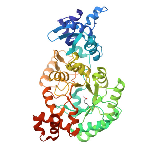
Experimental Data Snapshot

  • Method: X-RAY DIFFRACTION
  • Resolution: 2.66 Å
  • R-Value Free: 
    0.277 (Depositor), 0.277 (DCC) 
  • R-Value Work: 
    0.253 (Depositor), 0.253 (DCC) 
  • R-Value Observed: 
    0.253 (Depositor) 

Starting Model: in silico
View more details

wwPDB Validation   3D Report Full Report


Ligand Structure Quality Assessment 


This is version 1.0 of the entry. See complete history


Literature

Ligand-mediated emergence of higher-order protein complexity

Liu, A.K.Kaeser, B.Pereira, J.H.Taylor-Kearney, L.J.Hammel, M.Nogales, N.Adams, P.D.Shih, P.M.

To be published.

Macromolecules
Find similar proteins by:  (by identity cutoff)  |  3D Structure
Entity ID: 1
MoleculeChains Sequence LengthOrganismDetailsImage
ribulose-1,5-bisphosphate carboxylase
A, B, C, D, E
A, B, C, D, E, F, G, H, I, J
468Methanococcoides burtoniiMutation(s): 0 
Entity Groups  
Sequence Clusters30% Identity50% Identity70% Identity90% Identity95% Identity100% Identity
Sequence Annotations
Expand
  • Reference Sequence
Small Molecules
Ligands 2 Unique
IDChains Name / Formula / InChI Key2D Diagram3D Interactions
CAP (Subject of Investigation/LOI)
Query on CAP

Download Ideal Coordinates CCD File 
CA [auth G]
FA [auth H]
JA [auth I]
K [auth A]
MA [auth J]
CA [auth G],
FA [auth H],
JA [auth I],
K [auth A],
MA [auth J],
O [auth B],
R [auth C],
U [auth D],
W [auth E],
Z [auth F]
2-CARBOXYARABINITOL-1,5-DIPHOSPHATE
C6 H14 O13 P2
ITHCSGCUQDMYAI-ZMIZWQJLSA-N
MG (Subject of Investigation/LOI)
Query on MG

Download Ideal Coordinates CCD File 
AA [auth F]
BA [auth F]
DA [auth G]
EA [auth G]
GA [auth H]
AA [auth F],
BA [auth F],
DA [auth G],
EA [auth G],
GA [auth H],
HA [auth H],
IA [auth I],
KA [auth I],
L [auth A],
LA [auth J],
M [auth A],
N [auth A],
NA [auth J],
P [auth B],
Q [auth B],
S [auth C],
T [auth D],
V [auth D],
X [auth E],
Y [auth E]
MAGNESIUM ION
Mg
JLVVSXFLKOJNIY-UHFFFAOYSA-N
Modified Residues  1 Unique
IDChains TypeFormula2D DiagramParent
KCX
Query on KCX
A, B, C, D, E
A, B, C, D, E, F, G, H, I, J
L-PEPTIDE LINKINGC7 H14 N2 O4LYS
Experimental Data & Validation

Experimental Data

  • Method: X-RAY DIFFRACTION
  • Resolution: 2.66 Å
  • R-Value Free:  0.277 (Depositor), 0.277 (DCC) 
  • R-Value Work:  0.253 (Depositor), 0.253 (DCC) 
  • R-Value Observed: 0.253 (Depositor) 
Space Group: P 21 21 21
Unit Cell:
Length ( Å )Angle ( ˚ )
a = 80.844α = 90
b = 247.856β = 90
c = 266.093γ = 90
Software Package:
Software NamePurpose
PHENIXrefinement
xia2data reduction
xia2data scaling
PHASERphasing

Structure Validation

View Full Validation Report


Ligand Structure Quality Assessment 


Entry History & Funding Information

Deposition Data


Funding OrganizationLocationGrant Number
Department of Energy (DOE, United States)United States--

Revision History  (Full details and data files)

  • Version 1.0: 2025-11-19
    Type: Initial release