9DS1 | pdb_00009ds1

Crystal structure of 241_2F04 Fab in complex with H1 HA from A/California/04/2009(H1N1)


Experimental Data Snapshot

  • Method: X-RAY DIFFRACTION
  • Resolution: 2.40 Å
  • R-Value Free: 
    0.263 (Depositor), 0.264 (DCC) 
  • R-Value Work: 
    0.220 (Depositor), 0.222 (DCC) 
  • R-Value Observed: 
    0.222 (Depositor) 

Starting Model: experimental
View more details

wwPDB Validation   3D Report Full Report


Ligand Structure Quality Assessment 


This is version 1.0 of the entry. See complete history


Literature

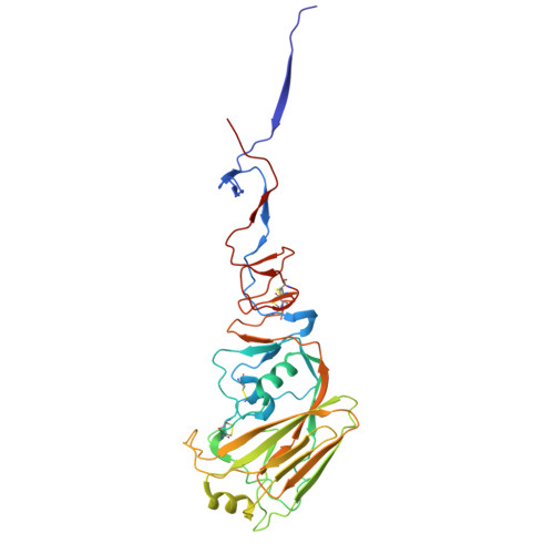
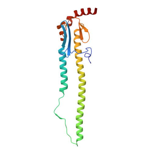
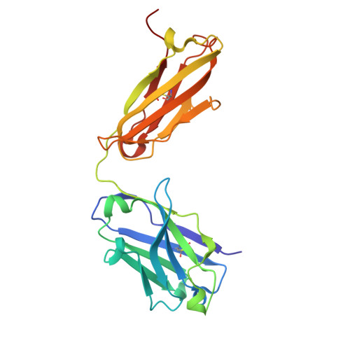
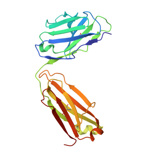
Structurally convergent antibodies derived from different vaccine strategies target the influenza virus HA anchor epitope with a subset of V H 3 and V K 3 genes.

Lin, T.H.Lee, C.D.Fernandez-Quintero, M.L.Ferguson, J.A.Han, J.Zhu, X.Yu, W.Guthmiller, J.J.Krammer, F.Wilson, P.C.Ward, A.B.Wilson, I.A.

(2025) Nat Commun 16: 1268-1268

  • DOI: https://doi.org/10.1038/s41467-025-56496-4
  • Primary Citation of Related Structures:  
    9DM0, 9DRU, 9DS1, 9DS2, 9DSC

  • PubMed Abstract: 

    H1N1 influenza viruses are responsible for both seasonal and pandemic influenza. The continual antigenic shift and drift of these viruses highlight the urgent need for a universal influenza vaccine to elicit broadly neutralizing antibodies (bnAbs). Identification and characterization of bnAbs elicited in natural infection and immunization to influenza virus hemagglutinin (HA) can provide insights for development of a universal influenza vaccine. Here, we structurally and biophysically characterize four antibodies that bind to a conserved region on the HA membrane-proximal region known as the anchor epitope. Despite some diversity in their V H and V K genes, the antibodies interact with the HA through germline-encoded residues in HCDR2 and LCDR3. Somatic mutations on HCDR3 also contribute hydrophobic interactions with the conserved HA epitope. This convergent binding mode provides extensive neutralization breadth against H1N1 viruses and suggests possible countermeasures against H1N1 viruses.


  • Organizational Affiliation
    • Department of Integrative Structural and Computational Biology, The Scripps Research Institute, La Jolla, CA, 92037, USA.

Macromolecules
Find similar proteins by:  (by identity cutoff)  |  3D Structure
Entity ID: 1
MoleculeChains Sequence LengthOrganismDetailsImage
Hemagglutinin HA1 chai327Influenza A virusMutation(s): 0 
Gene Names: HA
UniProt
Find proteins for C3W5S1 (Influenza A virus (strain swl A/California/04/2009 H1N1))
Explore C3W5S1 
Go to UniProtKB:  C3W5S1
Entity Groups  
Sequence Clusters30% Identity50% Identity70% Identity90% Identity95% Identity100% Identity
UniProt GroupC3W5S1
Glycosylation
Glycosylation Sites: 2
Sequence Annotations
Expand
  • Reference Sequence
Find similar proteins by:  (by identity cutoff)  |  3D Structure
Entity ID: 2
MoleculeChains Sequence LengthOrganismDetailsImage
Hemagglutinin HA2 chain175Influenza A virusMutation(s): 0 
Gene Names: HA
UniProt
Find proteins for E0UV84 (Influenza A virus)
Explore E0UV84 
Go to UniProtKB:  E0UV84
Entity Groups  
Sequence Clusters30% Identity50% Identity70% Identity90% Identity95% Identity100% Identity
UniProt GroupE0UV84
Sequence Annotations
Expand
  • Reference Sequence
Find similar proteins by:  (by identity cutoff)  |  3D Structure
Entity ID: 3
MoleculeChains Sequence LengthOrganismDetailsImage
241_2F04, Heavy chainC [auth H]226Homo sapiensMutation(s): 0 
Entity Groups  
Sequence Clusters30% Identity50% Identity70% Identity90% Identity95% Identity100% Identity
Sequence Annotations
Expand
  • Reference Sequence
Find similar proteins by:  (by identity cutoff)  |  3D Structure
Entity ID: 4
MoleculeChains Sequence LengthOrganismDetailsImage
241_2F04, Light chainD [auth L]215Homo sapiensMutation(s): 0 
Entity Groups  
Sequence Clusters30% Identity50% Identity70% Identity90% Identity95% Identity100% Identity
Sequence Annotations
Expand
  • Reference Sequence
Oligosaccharides

Help

Entity ID: 5
MoleculeChains Length2D Diagram Glycosylation3D Interactions
beta-D-mannopyranose-(1-4)-2-acetamido-2-deoxy-beta-D-glucopyranose-(1-4)-2-acetamido-2-deoxy-beta-D-glucopyranoseE [auth C]3N-Glycosylation
Glycosylation Resources
GlyTouCan:  G15407YE
GlyCosmos:  G15407YE
GlyGen:  G15407YE
Small Molecules
Ligands 1 Unique
IDChains Name / Formula / InChI Key2D Diagram3D Interactions
NAG (Subject of Investigation/LOI)
Query on NAG

Download Ideal Coordinates CCD File 
F [auth A]2-acetamido-2-deoxy-beta-D-glucopyranose
C8 H15 N O6
OVRNDRQMDRJTHS-FMDGEEDCSA-N
Experimental Data & Validation

Experimental Data

  • Method: X-RAY DIFFRACTION
  • Resolution: 2.40 Å
  • R-Value Free:  0.263 (Depositor), 0.264 (DCC) 
  • R-Value Work:  0.220 (Depositor), 0.222 (DCC) 
  • R-Value Observed: 0.222 (Depositor) 
Space Group: H 3 2
Unit Cell:
Length ( Å )Angle ( ˚ )
a = 129.466α = 90
b = 129.466β = 90
c = 443.769γ = 120
Software Package:
Software NamePurpose
PHENIXrefinement
HKL-2000data reduction
HKL-2000data scaling
PHASERphasing

Structure Validation

View Full Validation Report


Ligand Structure Quality Assessment 


Entry History & Funding Information

Deposition Data


Funding OrganizationLocationGrant Number
National Institutes of Health/National Institute Of Allergy and Infectious Diseases (NIH/NIAID)United States75N93019C00051

Revision History  (Full details and data files)

  • Version 1.0: 2025-03-19
    Type: Initial release