9DI0 | pdb_00009di0

Cryo-EM structure of Kif18A bound to a microtubule


Experimental Data Snapshot

  • Method: ELECTRON MICROSCOPY
  • Resolution: 3.10 Å
  • Aggregation State: HELICAL ARRAY 
  • Reconstruction Method: HELICAL 

wwPDB Validation 3D Report Full Report

Validation slider image for 9DI0

This is version 1.0 of the entry. See complete history

Literature

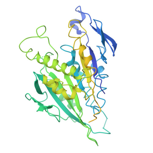
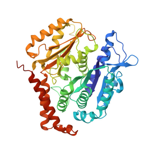
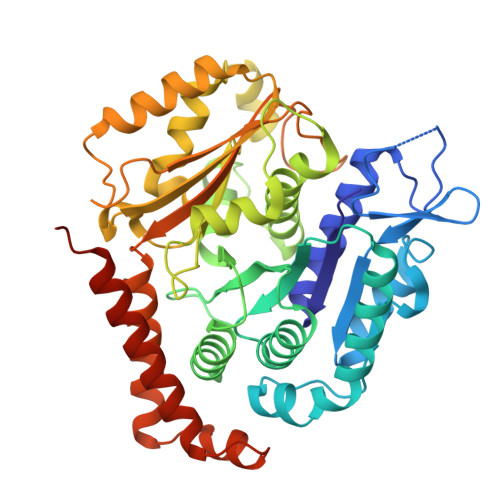
HURP regulates Kif18A recruitment and activity to synergistically control microtubule dynamics.

Perez-Bertoldi, J.M.Zhao, Y.Thawani, A.Yildiz, A.Nogales, E.

(2024) Nat Commun 15: 9687-9687

  • DOI: https://doi.org/10.1038/s41467-024-53691-7
  • Primary Citation Related Structures: 
    9DHZ, 9DI0, 9DXC, 9DXE

  • PubMed Abstract: 

    During mitosis, microtubule dynamics are regulated to ensure proper alignment and segregation of chromosomes. The dynamics of kinetochore-attached microtubules are regulated by hepatoma-upregulated protein (HURP) and the mitotic kinesin-8 Kif18A, but the underlying mechanism remains elusive. Using single-molecule imaging in vitro, we demonstrate that Kif18A motility is regulated by HURP. While sparse decoration of HURP activates the motor, higher concentrations hinder processive motility. To shed light on this behavior, we determine the binding mode of HURP to microtubules using cryo-EM. The structure helps rationalize why HURP functions as a microtubule stabilizer. Additionally, HURP partially overlaps with the microtubule-binding site of the Kif18A motor domain, indicating that excess HURP inhibits Kif18A motility by steric exclusion. We also observe that HURP and Kif18A function together to suppress dynamics of the microtubule plus-end, providing a mechanistic basis for how they collectively serve in microtubule length control.


  • Organizational Affiliation
    • Biophysics Graduate Group, University of California, Berkeley, CA, USA.

Macromolecule Content 

  • Total Structure Weight: 164.42 kDa 
  • Atom Count: 9,361 
  • Modeled Residue Count: 1,175 
  • Deposited Residue Count: 1,458 
  • Unique protein chains: 3

Macromolecules

Find similar proteins by:|  3D Structure
Entity ID: 1
MoleculeChains  Sequence LengthOrganismDetailsImage
Kinesin-like protein KIF18A, Methylated-DNA--protein-cysteine methyltransferase chimera562Homo sapiensMutation(s): 0 
Gene Names: KIF18AOK/SW-cl.108
EC: 2.1.1.63
UniProt & NIH Common Fund Data Resources
Find proteins for P16455 (Homo sapiens)
Explore P16455 
Go to UniProtKB:  P16455
PHAROS:  P16455
GTEx:  ENSG00000170430 
Find proteins for Q8NI77 (Homo sapiens)
Explore Q8NI77 
Go to UniProtKB:  Q8NI77
PHAROS:  Q8NI77
GTEx:  ENSG00000121621 
Entity Groups
Sequence Clusters30% Identity50% Identity70% Identity90% Identity95% Identity100% Identity
UniProt GroupsP16455Q8NI77
Sequence Annotations
Expand
Reference Sequence
Find similar proteins by:|  3D Structure
Entity ID: 2
MoleculeChains  Sequence LengthOrganismDetailsImage
Tubulin beta chainB [auth K]445Sus scrofaMutation(s): 0 
UniProt
Find proteins for P02554 (Sus scrofa)
Explore P02554 
Go to UniProtKB:  P02554
Entity Groups
Sequence Clusters30% Identity50% Identity70% Identity90% Identity95% Identity100% Identity
UniProt GroupP02554
Sequence Annotations
Expand
Reference Sequence
Find similar proteins by:|  3D Structure
Entity ID: 3
MoleculeChains  Sequence LengthOrganismDetailsImage
Tubulin alpha-1B chainC [auth L]451Sus scrofaMutation(s): 0 
EC: 3.6.5
UniProt
Find proteins for Q2XVP4 (Sus scrofa)
Explore Q2XVP4 
Go to UniProtKB:  Q2XVP4
Entity Groups
Sequence Clusters30% Identity50% Identity70% Identity90% Identity95% Identity100% Identity
UniProt GroupQ2XVP4
Sequence Annotations
Expand
Reference Sequence

Small Molecules

Ligands 5 Unique
IDChains Name / Formula / InChI Key2D Diagram3D Interactions
TA1

Query on TA1


Download:Ideal Coordinates CCD File
F [auth K]TAXOL
C47 H51 N O14
RCINICONZNJXQF-MZXODVADSA-N
GTP

Query on GTP


Download:Ideal Coordinates CCD File
H [auth L]GUANOSINE-5'-TRIPHOSPHATE
C10 H16 N5 O14 P3
XKMLYUALXHKNFT-UUOKFMHZSA-N
GDP

Query on GDP


Download:Ideal Coordinates CCD File
G [auth K]GUANOSINE-5'-DIPHOSPHATE
C10 H15 N5 O11 P2
QGWNDRXFNXRZMB-UUOKFMHZSA-N
ADP

Query on ADP


Download:Ideal Coordinates CCD File
D [auth A]ADENOSINE-5'-DIPHOSPHATE
C10 H15 N5 O10 P2
XTWYTFMLZFPYCI-KQYNXXCUSA-N
MG

Query on MG


Download:Ideal Coordinates CCD File
E [auth A],
I [auth L]
MAGNESIUM ION
Mg
JLVVSXFLKOJNIY-UHFFFAOYSA-N

Experimental Data & Validation

Experimental Data

  • Method: ELECTRON MICROSCOPY
  • Resolution: 3.10 Å
  • Aggregation State: HELICAL ARRAY 
  • Reconstruction Method: HELICAL 

Structure Validation

View Full Validation Report


Entry History 

& Funding Information

Deposition Data


Funding OrganizationLocationGrant Number
European Research Council (ERC)European Union--
Howard Hughes Medical Institute (HHMI)United States--
National Institutes of Health/National Institute of General Medical Sciences (NIH/NIGMS)United States--

Revision History  (Full details and data files)

  • Version 1.0: 2025-07-16
    Type: Initial release