9DDD | pdb_00009ddd

SufS-SufU complex from Mycobacterium Tuberculosis


Experimental Data Snapshot

  • Method: X-RAY DIFFRACTION
  • Resolution: 2.00 Å
  • R-Value Free: 
    0.203 (Depositor), 0.210 (DCC) 
  • R-Value Work: 
    0.179 (Depositor), 0.188 (DCC) 
  • R-Value Observed: 
    0.181 (Depositor) 

Starting Models: in silico
View more details

wwPDB Validation 3D Report Full Report

Validation slider image for 9DDD

Ligand Structure Quality Assessment 


This is version 1.0 of the entry. See complete history

Literature

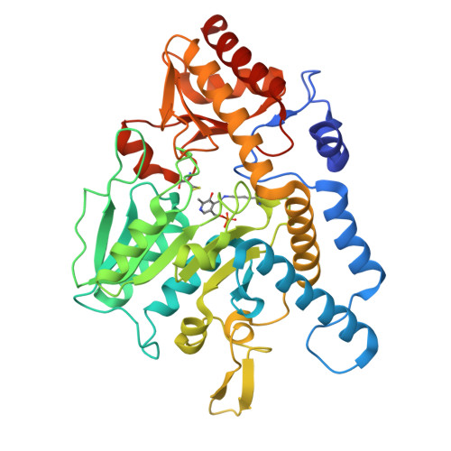
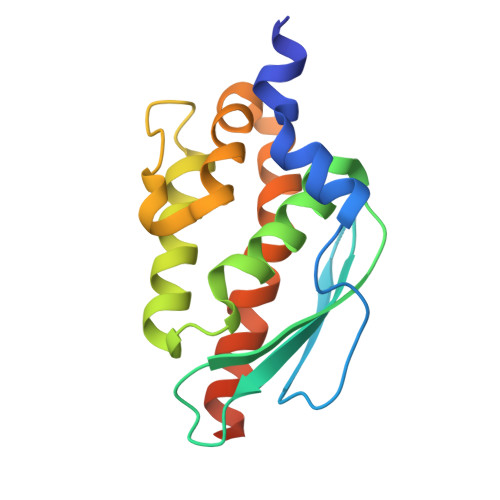
Feedback regulation of iron-sulfur cluster biogenesis.

Stuteley, S.M.Chen, J.Wang, J.Dawes, S.Baker, E.N.Squire, C.J.Pandelia, M.E.Bashiri, G.

(2025) bioRxiv 

  • DOI: https://doi.org/10.1101/2025.06.15.659787
  • Primary Citation Related Structures: 
    9DCL, 9DDD

  • PubMed Abstract: 

    Iron-sulfur (Fe-S) clusters are ubiquitous cofactors in biological systems. Given their central role in bacterial metabolism and pathogenesis, the biogenesis of Fe-S clusters is tightly controlled. We reveal a feedback regulatory mechanism involving the sulfide producing SufS/SufU complex within the sulfur utilization (SUF) system of Mycobacterium tuberculosis , the bacterium that causes tuberculosis. In this mechanism, [2Fe-2S] clusters compete with zinc ions for binding to the sulfide transfer protein SufU. Cluster binding induces SufU tetramerization, which prevents its interaction with the cysteine desulfurase SufS, thereby inhibiting SufS activation and limiting sulfide supply for Fe-S cluster biogenesis. These findings uncover an unrecognized regulatory mechanism in M. tuberculosis , ensuring strict control of Fe-S cluster production.

Macromolecule Content 

  • Total Structure Weight: 125.52 kDa 
  • Atom Count: 8,668 
  • Modeled Residue Count: 1,098 
  • Deposited Residue Count: 1,158 
  • Unique protein chains: 2

Macromolecules

Find similar proteins by:|  3D Structure
Entity ID: 1
MoleculeChains  Sequence LengthOrganismDetailsImage
Probable cysteine desulfurase
A, B
417Mycobacterium tuberculosisMutation(s): 0 
Gene Names: csdRv1464MTV007.11
EC: 2.8.1.7
UniProt
Find proteins for P9WQ69 (Mycobacterium tuberculosis (strain ATCC 25618 / H37Rv))
Explore P9WQ69 
Go to UniProtKB:  P9WQ69
Entity Groups
Sequence Clusters30% Identity50% Identity70% Identity90% Identity95% Identity100% Identity
UniProt GroupP9WQ69
Sequence Annotations
Expand
Reference Sequence
Find similar proteins by:|  3D Structure
Entity ID: 2
MoleculeChains  Sequence LengthOrganismDetailsImage
Possible nitrogen fixation related protein
C, D
162Mycobacterium tuberculosisMutation(s): 0 
Gene Names: Rv1465
UniProt
Find proteins for O53156 (Mycobacterium tuberculosis (strain ATCC 25618 / H37Rv))
Explore O53156 
Go to UniProtKB:  O53156
Entity Groups
Sequence Clusters30% Identity50% Identity70% Identity90% Identity95% Identity100% Identity
UniProt GroupO53156
Sequence Annotations
Expand
Reference Sequence

Experimental Data & Validation

Experimental Data

  • Method: X-RAY DIFFRACTION
  • Resolution: 2.00 Å
  • R-Value Free:  0.203 (Depositor), 0.210 (DCC) 
  • R-Value Work:  0.179 (Depositor), 0.188 (DCC) 
  • R-Value Observed: 0.181 (Depositor) 
Space Group: P 1
Unit Cell:
Length ( Å )Angle ( ˚ )
a = 66.799α = 91.03
b = 73.616β = 100.22
c = 76.349γ = 110.02
Software Package:
Software NamePurpose
REFMACrefinement
XDSdata reduction
Aimlessdata scaling
PHENIXphasing

Structure Validation

View Full Validation Report


Ligand Structure Quality Assessment 


Entry History 

& Funding Information

Deposition Data


Funding OrganizationLocationGrant Number
Health Research Council (HRC)New Zealand--

Revision History  (Full details and data files)

  • Version 1.0: 2025-07-30
    Type: Initial release