9D9Z | pdb_00009d9z

Structure of human UBR4-KCMF1-CaM E3 ligase complex (Silencing Factor of the Integrated stress response, SiFI)

  • Classification: LIGASE
  • Organism(s): Homo sapiens
  • Mutation(s): No 

  • Deposited: 2024-08-21 Released: 2025-06-11 
  • Deposition Author(s): Yang, Z., Rape, M.
  • Funding Organization(s): The G. Harold and Leila Y. Mathers Foundation, Howard Hughes Medical Institute (HHMI)

Experimental Data Snapshot

  • Method: ELECTRON MICROSCOPY
  • Resolution: 3.40 Å
  • Aggregation State: PARTICLE 
  • Reconstruction Method: SINGLE PARTICLE 

wwPDB Validation   3D Report Full Report


This is version 1.1 of the entry. See complete history


Literature

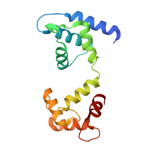
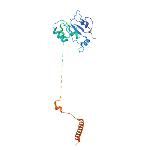
Molecular basis of SIFI activity in the integrated stress response.

Yang, Z.Haakonsen, D.L.Heider, M.Witus, S.R.Zelter, A.Beschauner, T.MacCoss, M.J.Rape, M.

(2025) Nature 643: 1117-1126

  • DOI: https://doi.org/10.1038/s41586-025-09074-z
  • Primary Citation of Related Structures:  
    9D9Z, 9NWD, 9NWE

  • PubMed Abstract: 

    Chronic stress response activation impairs cell survival and causes devastating degenerative diseases 1-3 . Organisms accordingly deploy silencing factors, such as the E3 ubiquitin ligase silencing factor of the integrated stress response (SIFI), to terminate stress response signalling and ensure cellular homeostasis 4 . How a silencing factor can sense stress across cellular scales to elicit timely stress response inactivation is poorly understood. Here we combine cryo-electron microscopy analysis of endogenous SIFI with AlphaFold modelling and biochemical studies to report the structural and mechanistic basis of the silencing of the integrated stress response. SIFI detects both stress indicators and stress response components through flexible domains within an easily accessible scaffold, before building linkage-specific ubiquitin chains at separate, sterically restricted elongation modules. Ubiquitin handover by a ubiquitin-like domain couples versatile substrate modification to linkage-specific ubiquitin polymer formation. Stress response silencing therefore exploits a catalytic mechanism that is geared towards processing many diverse proteins and therefore allows a single enzyme to monitor and, if needed, modulate a complex cellular state.


  • Organizational Affiliation
    • Department of Molecular and Cell Biology, University of California at Berkeley, Berkeley, CA, USA.

Macromolecules
Find similar proteins by:  (by identity cutoff)  |  3D Structure
Entity ID: 1
MoleculeChains Sequence LengthOrganismDetailsImage
UBR4 (endogenously FLAG-tagged at the N-terminus),E3 ubiquitin-protein ligase UBR4,E3 ubiquitin-protein ligase UBR4,E3 ubiquitin-protein ligase UBR4
A, B
5,205Homo sapiensMutation(s): 0 
EC: 2.3.2.27
UniProt & NIH Common Fund Data Resources
Find proteins for Q5T4S7 (Homo sapiens)
Explore Q5T4S7 
Go to UniProtKB:  Q5T4S7
PHAROS:  Q5T4S7
GTEx:  ENSG00000127481 
Entity Groups  
Sequence Clusters30% Identity50% Identity70% Identity90% Identity95% Identity100% Identity
UniProt GroupQ5T4S7
Sequence Annotations
Expand
  • Reference Sequence
Find similar proteins by:  (by identity cutoff)  |  3D Structure
Entity ID: 2
MoleculeChains Sequence LengthOrganismDetailsImage
Calmodulin-1
C, D
149Homo sapiensMutation(s): 0 
UniProt & NIH Common Fund Data Resources
Find proteins for P0DP23 (Homo sapiens)
Explore P0DP23 
Go to UniProtKB:  P0DP23
PHAROS:  P0DP23
GTEx:  ENSG00000198668 
Entity Groups  
Sequence Clusters30% Identity50% Identity70% Identity90% Identity95% Identity100% Identity
UniProt GroupP0DP23
Sequence Annotations
Expand
  • Reference Sequence
Find similar proteins by:  (by identity cutoff)  |  3D Structure
Entity ID: 3
MoleculeChains Sequence LengthOrganismDetailsImage
E3 ubiquitin-protein ligase KCMF1
E, F
381Homo sapiensMutation(s): 0 
EC: 2.3.2.27
UniProt & NIH Common Fund Data Resources
Find proteins for Q9P0J7 (Homo sapiens)
Explore Q9P0J7 
Go to UniProtKB:  Q9P0J7
PHAROS:  Q9P0J7
GTEx:  ENSG00000176407 
Entity Groups  
Sequence Clusters30% Identity50% Identity70% Identity90% Identity95% Identity100% Identity
UniProt GroupQ9P0J7
Sequence Annotations
Expand
  • Reference Sequence
Small Molecules
Ligands 2 Unique
IDChains Name / Formula / InChI Key2D Diagram3D Interactions
ZN
Query on ZN

Download Ideal Coordinates CCD File 
AA [auth F]
BA [auth F]
CA [auth F]
DA [auth F]
G [auth A]
AA [auth F],
BA [auth F],
CA [auth F],
DA [auth F],
G [auth A],
H [auth A],
I [auth A],
J [auth A],
K [auth A],
L [auth A],
M [auth B],
N [auth B],
O [auth B],
P [auth B],
Q [auth B],
R [auth B],
W [auth E],
X [auth E],
Y [auth E],
Z [auth E]
ZINC ION
Zn
PTFCDOFLOPIGGS-UHFFFAOYSA-N
CA
Query on CA

Download Ideal Coordinates CCD File 
S [auth C],
T [auth C],
U [auth D],
V [auth D]
CALCIUM ION
Ca
BHPQYMZQTOCNFJ-UHFFFAOYSA-N
Experimental Data & Validation

Experimental Data

  • Method: ELECTRON MICROSCOPY
  • Resolution: 3.40 Å
  • Aggregation State: PARTICLE 
  • Reconstruction Method: SINGLE PARTICLE 
EM Software:
TaskSoftware PackageVersion
RECONSTRUCTIONcryoSPARC4.3
MODEL REFINEMENTCoot
MODEL REFINEMENTPHENIX

Structure Validation

View Full Validation Report


Entry History & Funding Information

Deposition Data


Funding OrganizationLocationGrant Number
The G. Harold and Leila Y. Mathers FoundationUnited States--
Howard Hughes Medical Institute (HHMI)United States--

Revision History  (Full details and data files)

  • Version 1.0: 2025-06-11
    Type: Initial release
  • Version 1.1: 2025-08-06
    Changes: Data collection, Database references