9D8B | pdb_00009d8b

High-resolution crystal structure of Vibrio cholerae NFeoB in the GDP-bound form


Experimental Data Snapshot

  • Method: X-RAY DIFFRACTION
  • Resolution: 1.82 Å
  • R-Value Free: 
    0.246 (Depositor), 0.247 (DCC) 
  • R-Value Work: 
    0.203 (Depositor), 0.204 (DCC) 
  • R-Value Observed: 
    0.205 (Depositor) 

Starting Model: experimental
View more details

wwPDB Validation 3D Report Full Report

Validation slider image for 9D8B

Ligand Structure Quality Assessment 


This is version 2.1 of the entry. See complete history

Literature

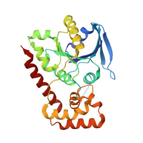
The Structure of the Full Catalytic Cycle of Vibrio cholerae NFeoB.

Magante, K.Armstrong, C.M.Lee, M.Smith, A.T.

(2026) J Mol Biology 438: 169727-169727

  • DOI: https://doi.org/10.1016/j.jmb.2026.169727
  • Primary Citation Related Structures: 
    9D8B, 9D8D

  • PubMed Abstract: 

    The acquisition of iron is critical for the survival and the virulence of numerous infectious pathogens, and most bacteria acquire ferrous iron (Fe 2+ ) by utilizing the ferrous iron transport (Feo) system. FeoB is the main component of this system and its function is regulated by its soluble cytosolic domain, termed NFeoB. We have recently begun to define the structure and the mechanism of the Feo system from the bacterium Vibrio cholerae, the causative agent of the disease cholera. However, major structural gaps in our understanding of the nucleotide-promiscuous V. cholerae NFeoB still exist. In this work, we have determined several new X-ray crystal structures that reveal distinct snapshots of the VcNFeoB domain in uncommon and unprecedented states, ultimately illuminating the full catalytic cycle of this NTPase. This work reveals important functional features of VcNFeoB that may be leveraged and ultimately targeted to prevent the infectivity and the spread of cholera.


  • Organizational Affiliation
    • Department of Chemistry and Biochemistry, University of Maryland, Baltimore County, Baltimore, MD 21250, USA.

Macromolecule Content 

  • Total Structure Weight: 119.82 kDa 
  • Atom Count: 8,205 
  • Modeled Residue Count: 988 
  • Deposited Residue Count: 1,044 
  • Unique protein chains: 1

Macromolecules

Find similar proteins by:|  3D Structure
Entity ID: 1
MoleculeChains  Sequence LengthOrganismDetailsImage
Ferrous iron transport protein B
A, B, C, D
261Vibrio choleraeMutation(s): 0 
Gene Names: feoB_1ERS013165_00117
UniProt
Find proteins for A0A0X1KYD0 (Vibrio cholerae (strain MO10))
Explore A0A0X1KYD0 
Go to UniProtKB:  A0A0X1KYD0
Entity Groups
Sequence Clusters30% Identity50% Identity70% Identity90% Identity95% Identity100% Identity
UniProt GroupA0A0X1KYD0
Sequence Annotations
Expand
Reference Sequence

Small Molecules

Ligands 2 Unique
IDChains Name / Formula / InChI Key2D Diagram3D Interactions
GDP

Query on GDP


Download:Ideal Coordinates CCD File
E [auth A],
H [auth B],
J [auth C],
N [auth D]
GUANOSINE-5'-DIPHOSPHATE
C10 H15 N5 O11 P2
QGWNDRXFNXRZMB-UUOKFMHZSA-N
GOL

Query on GOL


Download:Ideal Coordinates CCD File
F [auth A]
G [auth A]
I [auth B]
K [auth C]
L [auth C]
F [auth A],
G [auth A],
I [auth B],
K [auth C],
L [auth C],
M [auth C],
O [auth D]
GLYCEROL
C3 H8 O3
PEDCQBHIVMGVHV-UHFFFAOYSA-N

Experimental Data & Validation

Experimental Data

  • Method: X-RAY DIFFRACTION
  • Resolution: 1.82 Å
  • R-Value Free:  0.246 (Depositor), 0.247 (DCC) 
  • R-Value Work:  0.203 (Depositor), 0.204 (DCC) 
  • R-Value Observed: 0.205 (Depositor) 
Space Group: P 1
Unit Cell:
Length ( Å )Angle ( ˚ )
a = 54.046α = 94.39
b = 59.024β = 92.98
c = 89.481γ = 113.21
Software Package:
Software NamePurpose
PHENIXrefinement
autoPROCdata reduction
autoPROCdata scaling
PHASERphasing

Structure Validation

View Full Validation Report


Ligand Structure Quality Assessment 


Entry History 

& Funding Information

Deposition Data


Funding OrganizationLocationGrant Number
National Institutes of Health/National Institute of General Medical Sciences (NIH/NIGMS)United StatesGM133497
National Science Foundation (NSF, United States)United StatesCHE1844624

Revision History  (Full details and data files)

  • Version 1.0: 2025-09-03
    Type: Initial release
  • Version 2.0: 2026-01-14
    Type: Coordinate replacement
    Reason: Polymer geometry
    Changes: Atomic model, Data collection, Derived calculations, Other, Refinement description, Structure summary
  • Version 2.1: 2026-03-18
    Changes: Database references