9D7W | pdb_00009d7w

The TBZ-bound structure


Experimental Data Snapshot

  • Method: ELECTRON MICROSCOPY
  • Resolution: 3.30 Å
  • Aggregation State: PARTICLE 
  • Reconstruction Method: SINGLE PARTICLE 

wwPDB Validation   3D Report Full Report


This is version 1.0 of the entry. See complete history


Literature

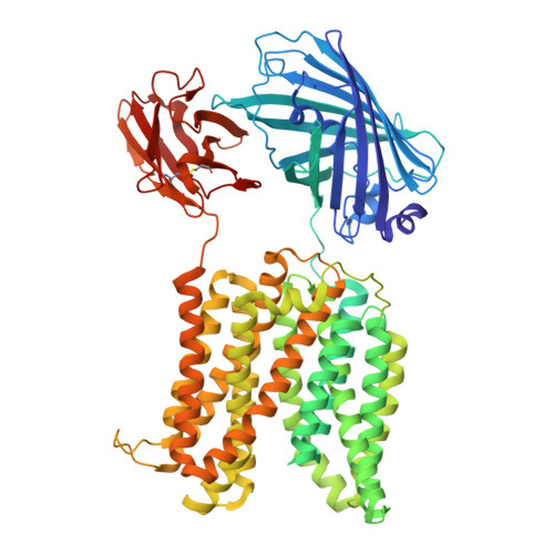
Structure and mechanism of human vesicular polyamine transporter.

Guo, Y.Yang, G.Liu, H.Chai, J.Chen, J.Shanklin, J.Liu, Q.Liu, B.Lu, M.

(2025) Nat Commun 16: 4142-4142

  • DOI: https://doi.org/10.1038/s41467-025-59549-w
  • Primary Citation of Related Structures:  
    9D7U, 9D7V, 9D7W, 9D7X

  • PubMed Abstract: 

    Polyamines play essential roles in gene expression and modulate neuronal transmission in mammals. Vesicular polyamine transporters (VPAT) from the SLC18 family exploit the transmembrane H + gradient to translocate polyamines into secretory vesicles, enabling the quantal release of polyamine neuromodulators and underpinning learning and memory formation. Here, we report the cryo-electron microscopy structures of human VPAT in complex with spermine, spermidine, H + , or tetrabenazine, elucidating discrete lumen-facing states of the antiporter and pivotal interactions between VPAT and its substrate or inhibitor. Leveraging structure-inspired mutagenesis studies and protein structure prediction, we deduce an unforeseen mechanism whereby the polyamine and H + compete for multiple acidic protein residues both directly and indirectly, and rationalize how the antidopaminergic therapeutic tetrabenazine impedes vesicular transport of polyamines. This study unravels the mechanism of an H + -coupled polyamine antiporter, reveals mechanistic diversity between VPAT and other SLC18 antiporters, and raises new prospects for combating human disorders of polyamine homeostasis.


  • Organizational Affiliation
    • Center for Proteomics & Molecular Therapeutics, Rosalind Franklin University of Medicine & Science, 3333 Green Bay Road, North Chicago, IL, 60064, USA.

Macromolecules
Find similar proteins by:  (by identity cutoff)  |  3D Structure
Entity ID: 1
MoleculeChains Sequence LengthOrganismDetailsImage
Green fluorescence protein,MFS-type transporter SLC18B1,membrane protein with TBZ751Homo sapiensMutation(s): 0 
Gene Names: APY04_3197SLC18B1C6orf192
Membrane Entity: Yes 
UniProt & NIH Common Fund Data Resources
Find proteins for A0A125NTU3 (Hyphomicrobium sulfonivorans)
Explore A0A125NTU3 
Go to UniProtKB:  A0A125NTU3
Find proteins for Q6NT16 (Homo sapiens)
Explore Q6NT16 
Go to UniProtKB:  Q6NT16
PHAROS:  Q6NT16
GTEx:  ENSG00000146409 
Entity Groups  
Sequence Clusters30% Identity50% Identity70% Identity90% Identity95% Identity100% Identity
UniProt GroupsQ6NT16A0A125NTU3
Sequence Annotations
Expand
  • Reference Sequence
Small Molecules
Ligands 1 Unique
IDChains Name / Formula / InChI Key2D Diagram3D Interactions
YHL
Query on YHL

Download Ideal Coordinates CCD File 
B [auth A]tetrabenazine
C19 H27 N O3
MKJIEFSOBYUXJB-GDBMZVCRSA-N
Experimental Data & Validation

Experimental Data

  • Method: ELECTRON MICROSCOPY
  • Resolution: 3.30 Å
  • Aggregation State: PARTICLE 
  • Reconstruction Method: SINGLE PARTICLE 
EM Software:
TaskSoftware PackageVersion
MODEL REFINEMENTPHENIX1.21_5207:

Structure Validation

View Full Validation Report


Entry History & Funding Information

Deposition Data

  • Released Date: 2025-06-25 
  • Deposition Author(s): Lu, M., Liu, B.

Funding OrganizationLocationGrant Number
National Institutes of Health/National Institute of General Medical Sciences (NIH/NIGMS)United StatesR01GM145642

Revision History  (Full details and data files)

  • Version 1.0: 2025-06-25
    Type: Initial release