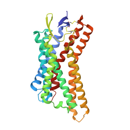
9CIB | pdb_00009cib

CryoEM Structure of HCA2 DREADD Gi1 in complex with FCH-2296413 (Local Refinement)


Experimental Data Snapshot

  • Method: ELECTRON MICROSCOPY
  • Resolution: 2.62 Å
  • Aggregation State: PARTICLE 
  • Reconstruction Method: SINGLE PARTICLE 

wwPDB Validation 3D Report Full Report

Validation slider image for 9CIB

This is version 1.0 of the entry. See complete history. 

Literature

Structure-guided design of a peripherally restricted chemogenetic system.

Kang, H.J., Krumm, B.E., Tassou, A., Geron, M., DiBerto, J.F., Kapolka, N.J., Gumpper, R.H., Sakamoto, K., Dewran Kocak, D., Olsen, R.H.J., Huang, X.P., Zhang, S., Huang, K.L., Zaidi, S.A., Nguyen, M.T., Jo, M.J., Katritch, V., Fay, J.F., Scherrer, G., Roth, B.L.

(2024) Cell 187: 7433-7449.e20

  • DOI: https://doi.org/10.1016/j.cell.2024.11.001
  • Primary Citation Related Structures: 
    8UTD, 8UUJ, 9CIB

  • PubMed Abstract: 

    Designer receptors exclusively activated by designer drugs (DREADDs) are chemogenetic tools for remotely controlling cellular signaling, neural activity, behavior, and physiology. Using a structure-guided approach, we provide a peripherally restricted Gi-DREADD, hydroxycarboxylic acid receptor DREADD (HCAD), whose native receptor is minimally expressed in the brain, and a chemical actuator that does not cross the blood-brain barrier (BBB). This was accomplished by combined mutagenesis, analoging via an ultra-large make-on-demand library, structural determination of the designed DREADD receptor via cryoelectron microscopy (cryo-EM), and validation of HCAD function. Expression and activation of HCAD in dorsal root ganglion (DRG) neurons inhibit action potential (AP) firing and reduce both acute and tissue-injury-induced inflammatory pain. The HCAD chemogenetic system expands the possibilities for studying numerous peripheral systems with little adverse effects on the central nervous system (CNS). The structure-guided approach used to generate HCAD also has the potential to accelerate the development of emerging chemogenetic tools for basic and translational sciences.


  • Organizational Affiliation: 
    • Department of Biotechnology, College of Life Science and Biotechnology, Yonsei University, Seoul, South Korea.

Macromolecule Content 

  • Total Structure Weight: 34.44 kDa 
  • Atom Count: 2,368 
  • Modeled Residue Count: 290 
  • Deposited Residue Count: 294 
  • Unique protein chains: 1

Macromolecules

Find similar proteins by:|  3D Structure
Entity ID: 1
MoleculeChains  Sequence LengthOrganismDetailsImage
Hydroxycarboxylic acid receptor 2A [auth R]294Mus musculusMutation(s): 2 
Gene Names: Hcar2, Gpr109, Gpr109a, Gpr109b, Niacr1, Pumag
Membrane Entity: Yes 
UniProt & NIH Common Fund Data Resources
Find proteins for Q9EP66 (Mus musculus)
Explore Q9EP66 
Go to UniProtKB:  Q9EP66
IMPC:  MGI:1933383
Entity Groups
Sequence Clusters30% Identity50% Identity70% Identity90% Identity95% Identity100% Identity
UniProt GroupQ9EP66
Sequence Annotations
Expand
Reference Sequence

Small Molecules

Ligands 1 Unique
IDChains Name / Formula / InChI Key2D Diagram3D Interactions
XI9
(Subject of Investigation/LOI)

Query on XI9


Download:Ideal Coordinates CCD File
B [auth R](3M,4aR,5aR)-3-(1H-tetrazol-5-yl)-4,4a,5,5a-tetrahydro-1H-cyclopropa[4,5]cyclopenta[1,2-c]pyrazole
C8 H8 N6
DKSPRNJUDCWTID-QWWZWVQMSA-N

Experimental Data & Validation

Experimental Data

  • Method: ELECTRON MICROSCOPY
  • Resolution: 2.62 Å
  • Aggregation State: PARTICLE 
  • Reconstruction Method: SINGLE PARTICLE 
EM Software:
TaskSoftware PackageVersion
MODEL REFINEMENTPHENIX

Structure Validation

View Full Validation Report


Entry History 

& Funding Information

Deposition Data


Funding OrganizationLocationGrant Number
National Institutes of Health/National Institute of Mental Health (NIH/NIMH)United States5R01MH112205

Revision History  (Full details and data files)

  • Version 1.0: 2025-05-14
    Type: Initial release