9C7E | pdb_00009c7e

Diheteromeric GluN1/GluN2A (delM653) in nanodisc complex with glycine, glutamate, and GNE-4123, open conformation, C2 symmetry


Experimental Data Snapshot

  • Method: ELECTRON MICROSCOPY
  • Resolution: 3.22 Å
  • Aggregation State: PARTICLE 
  • Reconstruction Method: SINGLE PARTICLE 

wwPDB Validation   3D Report Full Report


This is version 1.1 of the entry. See complete history


Literature

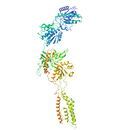
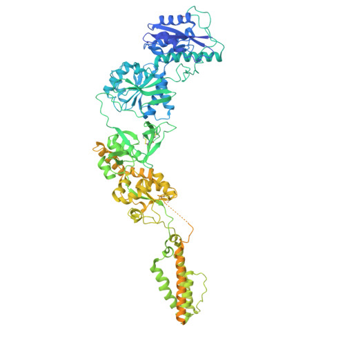
Cryo-EM snapshots of NMDA receptor activation illuminate sequential rearrangements.

Abbott, J.A.Kim, J.Liu, B.Popescu, G.K.Gouaux, E.Jalali-Yazdi, F.

(2025) Sci Adv 11: eadx4647-eadx4647

  • DOI: https://doi.org/10.1126/sciadv.adx4647
  • Primary Citation of Related Structures:  
    9C7C, 9C7E, 9C7P, 9C7Q, 9C7R

  • PubMed Abstract: 

    Canonical N -methyl-d-aspartate receptors (NMDARs) are glutamate-gated ion channels with critical roles in the development and function of the nervous system. The excitatory currents they produce reflect stochastic transitions between multiple agonist-bound closed- and open-pore states. We leveraged the intrinsically high open probability ( P o ) of NMDARs composed of GluN1 and GluN2A subunits, together with judiciously chosen mutants and ligands, to achieve conditions in which receptors had a P o near unity. Using single-particle cryo-electron microscopy (cryo-EM), we captured three activated receptor states, each with distinct conformations of the gate-forming M3 helices. Separately, we carried out single-channel electrophysiology, together with statistical modeling, to relate the cryo-EM structures to the gating reaction. NMDAR channel opening involves bending of the pore-forming M3 helices to produce a transient open-channel conformation, subsequently stabilized by new interactions between the D2-M3 linkers with the pre-M1 helices and the pre-M4 loops, to yield the stable open channel.


  • Organizational Affiliation
    • Department of Biochemistry, Jacobs School of Medicine and Biomedical Sciences, University at Buffalo, SUNY, Buffalo, NY 14203, USA.

Macromolecules
Find similar proteins by:  (by identity cutoff)  |  3D Structure
Entity ID: 1
MoleculeChains Sequence LengthOrganismDetailsImage
Glutamate receptor ionotropic, NMDA 1,Green fluorescent protein chimera
A, C
1,108Rattus norvegicusAequorea victoria
This entity is chimeric
Mutation(s): 0 
Gene Names: Grin1Nmdar1GFP
UniProt
Find proteins for P35439 (Rattus norvegicus)
Explore P35439 
Go to UniProtKB:  P35439
Find proteins for P42212 (Aequorea victoria)
Explore P42212 
Go to UniProtKB:  P42212
Entity Groups  
Sequence Clusters30% Identity50% Identity70% Identity90% Identity95% Identity100% Identity
UniProt GroupsP42212P35439
Sequence Annotations
Expand
  • Reference Sequence
Find similar proteins by:  (by identity cutoff)  |  3D Structure
Entity ID: 2
MoleculeChains Sequence LengthOrganismDetailsImage
Glutamate receptor ionotropic, NMDA 2A,Green fluorescent protein chimera
B, D
1,126Rattus norvegicusAequorea victoria
This entity is chimeric
Mutation(s): 0 
Gene Names: Grin2aGFP
UniProt
Find proteins for Q00959 (Rattus norvegicus)
Explore Q00959 
Go to UniProtKB:  Q00959
Find proteins for P42212 (Aequorea victoria)
Explore P42212 
Go to UniProtKB:  P42212
Entity Groups  
Sequence Clusters30% Identity50% Identity70% Identity90% Identity95% Identity100% Identity
UniProt GroupsQ00959P42212
Sequence Annotations
Expand
  • Reference Sequence
Small Molecules
Ligands 3 Unique
IDChains Name / Formula / InChI Key2D Diagram3D Interactions
Y01 (Subject of Investigation/LOI)
Query on Y01

Download Ideal Coordinates CCD File 
F [auth B]
G [auth B]
I [auth B]
K [auth D]
L [auth D]
F [auth B],
G [auth B],
I [auth B],
K [auth D],
L [auth D],
N [auth D]
CHOLESTEROL HEMISUCCINATE
C31 H50 O4
WLNARFZDISHUGS-MIXBDBMTSA-N
A1AUV (Subject of Investigation/LOI)
Query on A1AUV

Download Ideal Coordinates CCD File 
E [auth A],
J [auth C]
4-cyclohexyl-N-[(8R)-2-cyclopropyl-7-hydroxy-5-methyl[1,2,4]triazolo[1,5-a]pyrimidin-6-yl]benzene-1-sulfonamide
C21 H25 N5 O3 S
NVBBYGHRHGFNKE-UHFFFAOYSA-N
D12
Query on D12

Download Ideal Coordinates CCD File 
H [auth B],
M [auth D]
DODECANE
C12 H26
SNRUBQQJIBEYMU-UHFFFAOYSA-N
Experimental Data & Validation

Experimental Data

  • Method: ELECTRON MICROSCOPY
  • Resolution: 3.22 Å
  • Aggregation State: PARTICLE 
  • Reconstruction Method: SINGLE PARTICLE 
EM Software:
TaskSoftware PackageVersion
MODEL REFINEMENTPHENIX
RECONSTRUCTIONcryoSPARC

Structure Validation

View Full Validation Report


Entry History & Funding Information

Deposition Data


Funding OrganizationLocationGrant Number
National Institutes of Health/National Institute of Neurological Disorders and Stroke (NIH/NINDS)United States--

Revision History  (Full details and data files)

  • Version 1.0: 2025-09-24
    Type: Initial release
  • Version 1.1: 2026-02-04
    Changes: Database references