Similar but Distinct-Biochemical Characterization of the Staphylococcus aureus Serine Hydrolases FphH and FphI.
Fellner, M., Randall, G., Bitac, I.R.C.G., Warrender, A.K., Sethi, A., Jelinek, R., Kass, I.(2025) Proteins 93: 1009-1021
- PubMed: 39726198 Search on PubMed
- DOI: https://doi.org/10.1002/prot.26785
- Primary Citation Related Structures:
8G0N, 9C0L, 9C0M, 9C0N - PubMed Abstract:
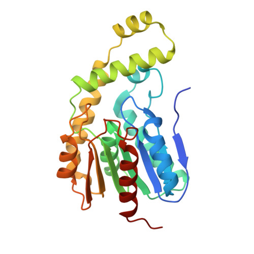
Staphylococcus aureus is a major cause of infections like bacteremia, pneumonia, and endocarditis. These infections are often linked to the ability of S. aureus to form biofilms. Several S. aureus serine hydrolases have previously been identified to be active during biofilm-forming conditions. Here, we present the biochemical characterization of two of these enzymes-fluorophosphonate binding hydrolase H and I (FphH, FphI). Cryogenic and room-temperature X-ray crystallography, enzymatic substrate profiling, small-angle X-ray scattering analysis, and molecular dynamics simulations provide new insights into similarities and differences between these two hydrolase_4 domain family members. We discover that these enzymes share an overall fold, including a flexible lid or cap region above the active site, which can be seen to be mobile in solution. Differences in the active site pocket and lid residues differentiate them and explain speed differences in their carboxyesterase substrate profile toward small unbranched carbon chain ester molecules. The first analysis of FphI is also compared to our previous knowledge of FphH and its association to stress conditions. These results enable the future precise targeting of Fph serine hydrolase family members with a long-term goal to significantly improve the health and wellbeing of individuals and populations worldwide.
- Biochemistry Department, School of Biomedical Sciences, University of Otago, Dunedin, New Zealand.
Organizational Affiliation: