


Crystal structure of Cry1Fa R226Q
Poland, B.W., Kakani, K., Yamamoto, T.To be published.
Experimental Data Snapshot
Starting Model: experimental
View more details
wwPDB Validation 3D Report Full Report

Entity ID: 1 | |||||
|---|---|---|---|---|---|
| Molecule | Chains | Sequence Length | Organism | Details | Image |
| Pesticidal crystal protein Cry1Fa | 1,174 | Bacillus thuringiensis | Mutation(s): 1 Gene Names: cry1Fa, cryIF, cryIF(a) | 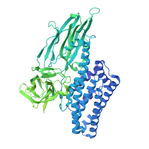 | |
UniProt | |||||
Entity Groups | |||||
| Sequence Clusters | 30% Identity50% Identity70% Identity90% Identity95% Identity100% Identity | ||||
| UniProt Group | Q03746 | ||||
Sequence AnnotationsExpand | |||||
Reference Sequence | |||||
| Length ( Å ) | Angle ( ˚ ) |
|---|---|
| a = 64.23 | α = 90 |
| b = 68.35 | β = 90 |
| c = 121.81 | γ = 90 |
| Software Name | Purpose |
|---|---|
| REFMAC | refinement |
| MOSFLM | data reduction |
| SCALA | data scaling |
| MOLREP | phasing |
| Funding Organization | Location | Grant Number |
|---|---|---|
| Not funded | United States | -- |


