9BTV | pdb_00009btv

Cryo-EM structure of rhesus antibody T646-a.01 in complex with HIV-1 Env trimer Q23.17 MD39


Experimental Data Snapshot

  • Method: ELECTRON MICROSCOPY
  • Resolution: 3.48 Å
  • Aggregation State: PARTICLE 
  • Reconstruction Method: SINGLE PARTICLE 

wwPDB Validation 3D Report Full Report

Validation slider image for 9BTV

This is version 1.1 of the entry. See complete history. 

Literature

Structural and genetic basis of HIV-1 envelope V2 apex recognition by rhesus broadly neutralizing antibodies.

Roark, R.S., Habib, R., Gorman, J., Li, H., Connell, A.J., Bonsignori, M., Guo, Y., Hogarty, M.P., Olia, A.S., Sowers, K.J., Zhang, B., Bibollet-Ruche, F., Bylund, T., Callaghan, S., Carey, J.W., Cerutti, G., Harris, D.R., He, W., Lewis, E., Liu, T., Mason, R.D., Qiao, Y., Park, Y., Rando, J.M., Singh, A., Wolff, J.J., Lei, Q.P., Louder, M.K., Andrabi, R., Doria-Rose, N.A., Saunders, K.O., Seaman, M.S., Haynes, B.F., Kulp, D.W., Mascola, J.R., Roederer, M., Pierson, T.C., Sheng, Z., Hahn, B.H., Shaw, G.M., Kwong, P.D., Shapiro, L.

(2025) J Exp Med 222

  • DOI: https://doi.org/10.1084/jem.20250638
  • Primary Citation Related Structures: 
    9BNK, 9BNL, 9BNM, 9BNP, 9BTH, 9BTI, 9BTJ, 9BTL, 9BTV

  • PubMed Abstract: 

    Broadly neutralizing antibodies targeting the V2 apex of HIV-1 envelope are desired as vaccine design templates, but few have been described. Here, we report 11 lineages of V2 apex-neutralizing antibodies from simian-human immunodeficiency virus (SHIV)-infected rhesus macaques and determine cryo-EM structures for 9. A single V2 apex-neutralizing lineage accounted for cross-clade breadth in most macaques, and somatic hypermutation relative to breadth was generally low, exemplified by antibody V033-a.01 with <5% nucleotide mutation and 37% breadth (208-strain panel). Envelope complex structures revealed eight different antibody classes (one multi-donor) and the complete repertoire of all five possible recognition topologies, recapitulating canonical human modes of apex insertion and C-strand hydrogen bonding. Despite this diversity in recognition, all rhesus-V2 apex antibodies were derived from reading frame two of the DH3-15*01 gene. Collectively, these results define-in rhesus-the structural and genetic basis of HIV-1 V2 apex recognition and demonstrate unprecedented structural plasticity of a highly selected immunogenetic element.


  • Organizational Affiliation: 

    • Aaron Diamond AIDS Research Center, Columbia University Vagelos College of Physicians and Surgeons , New York, NY, USA.

Macromolecule Content 

  • Total Structure Weight: 285.92 kDa 
  • Atom Count: 16,852 
  • Modeled Residue Count: 1,915 
  • Deposited Residue Count: 2,327 
  • Unique protein chains: 4

Macromolecules

Find similar proteins by:|  3D Structure
Entity ID: 1
MoleculeChains  Sequence LengthOrganismDetailsImage
Envelope glycoprotein gp120A,
C,
D [auth E]
469Human immunodeficiency virus 1Mutation(s): 0 
Gene Names: env
UniProt
Find proteins for O55774 (Human immunodeficiency virus type 1)
Explore O55774 
Go to UniProtKB:  O55774
Entity Groups
Sequence Clusters30% Identity50% Identity70% Identity90% Identity95% Identity100% Identity
UniProt GroupO55774
Glycosylation
Glycosylation Sites: 17
Sequence Annotations
Expand
Reference Sequence
Find similar proteins by:|  3D Structure
Entity ID: 2
MoleculeChains  Sequence LengthOrganismDetailsImage
Q23.MD39 Transmembrane protein gp41B,
E [auth F],
F [auth G]
153Human immunodeficiency virus 1Mutation(s): 7 
Gene Names: env
UniProt
Find proteins for O55774 (Human immunodeficiency virus type 1)
Explore O55774 
Go to UniProtKB:  O55774
Entity Groups
Sequence Clusters30% Identity50% Identity70% Identity90% Identity95% Identity100% Identity
UniProt GroupO55774
Glycosylation
Glycosylation Sites: 3
Sequence Annotations
Expand
Reference Sequence
Find similar proteins by:|  3D Structure
Entity ID: 3
MoleculeChains  Sequence LengthOrganismDetailsImage
T646-a.01 heavy chainG [auth H]245Macaca mulattaMutation(s): 0 
Entity Groups
Sequence Clusters30% Identity50% Identity70% Identity90% Identity95% Identity100% Identity
Sequence Annotations
Expand
Reference Sequence
Find similar proteins by:|  3D Structure
Entity ID: 4
MoleculeChains  Sequence LengthOrganismDetailsImage
T646-a.01 light chainH [auth L]216Macaca mulattaMutation(s): 0 
Entity Groups
Sequence Clusters30% Identity50% Identity70% Identity90% Identity95% Identity100% Identity
Sequence Annotations
Expand
Reference Sequence

Oligosaccharides

Help  
Entity ID: 5
MoleculeChains Length2D Diagram GlycosylationD Interactions
alpha-D-mannopyranose-(1-3)-[alpha-D-mannopyranose-(1-6)]beta-D-mannopyranose-(1-4)-2-acetamido-2-deoxy-beta-D-glucopyranose-(1-4)-2-acetamido-2-deoxy-beta-D-glucopyranoseI [auth D]5N-Glycosylation
Glycosylation Resources
GlyTouCan: G22768VO
GlyCosmos: G22768VO
GlyGen: G22768VO
Entity ID: 6
MoleculeChains Length2D Diagram GlycosylationD Interactions
alpha-D-mannopyranose-(1-3)-beta-D-mannopyranose-(1-4)-2-acetamido-2-deoxy-beta-D-glucopyranose-(1-4)-2-acetamido-2-deoxy-beta-D-glucopyranoseJ [auth I]4N-Glycosylation
Glycosylation Resources
GlyTouCan: G81315DD
GlyCosmos: G81315DD
GlyGen: G81315DD
Entity ID: 7
MoleculeChains Length2D Diagram GlycosylationD Interactions
beta-D-mannopyranose-(1-4)-2-acetamido-2-deoxy-beta-D-glucopyranose-(1-4)-2-acetamido-2-deoxy-beta-D-glucopyranose
DA [auth d],
GA [auth g],
K [auth J],
L [auth K],
MA [auth m],
DA [auth d],
GA [auth g],
K [auth J],
L [auth K],
MA [auth m],
NA [auth n],
Q,
RA [auth r],
T,
UA [auth u],
X,
Y
3N-Glycosylation
Glycosylation Resources
GlyTouCan: G15407YE
GlyCosmos: G15407YE
GlyGen: G15407YE
Entity ID: 9
MoleculeChains Length2D Diagram GlycosylationD Interactions
alpha-D-mannopyranose-(1-6)-beta-D-mannopyranose-(1-4)-2-acetamido-2-deoxy-beta-D-glucopyranose-(1-4)-2-acetamido-2-deoxy-beta-D-glucopyranoseIA [auth i],
KA [auth k]
4N-Glycosylation
Glycosylation Resources
GlyTouCan: G22573RC
GlyCosmos: G22573RC
GlyGen: G22573RC
Entity ID: 10
MoleculeChains Length2D Diagram GlycosylationD Interactions
alpha-D-mannopyranose-(1-6)-alpha-D-mannopyranose-(1-6)-[alpha-D-mannopyranose-(1-3)]beta-D-mannopyranose-(1-4)-2-acetamido-2-deoxy-beta-D-glucopyranose-(1-4)-2-acetamido-2-deoxy-beta-D-glucopyranoseJA [auth j]6N-Glycosylation
Glycosylation Resources
GlyTouCan: G34442SS
GlyCosmos: G34442SS
GlyGen: G34442SS

Small Molecules

Ligands 1 Unique
IDChains Name / Formula / InChI Key2D Diagram3D Interactions
NAG

Query on NAG


Download:Ideal Coordinates CCD File
AB [auth A]
BB [auth B]
CB [auth B]
DB [auth C]
EB [auth C]
AB [auth A],
BB [auth B],
CB [auth B],
DB [auth C],
EB [auth C],
FB [auth C],
GB [auth C],
HB [auth E],
IB [auth E],
JB [auth E],
KB [auth E],
LB [auth F],
MB [auth F],
NB [auth G],
OB [auth G],
PB [auth G],
XA [auth A],
YA [auth A],
ZA [auth A]
2-acetamido-2-deoxy-beta-D-glucopyranose
C8 H15 N O6
OVRNDRQMDRJTHS-FMDGEEDCSA-N
Modified Residues  1 Unique
IDChains TypeFormula2D DiagramParent
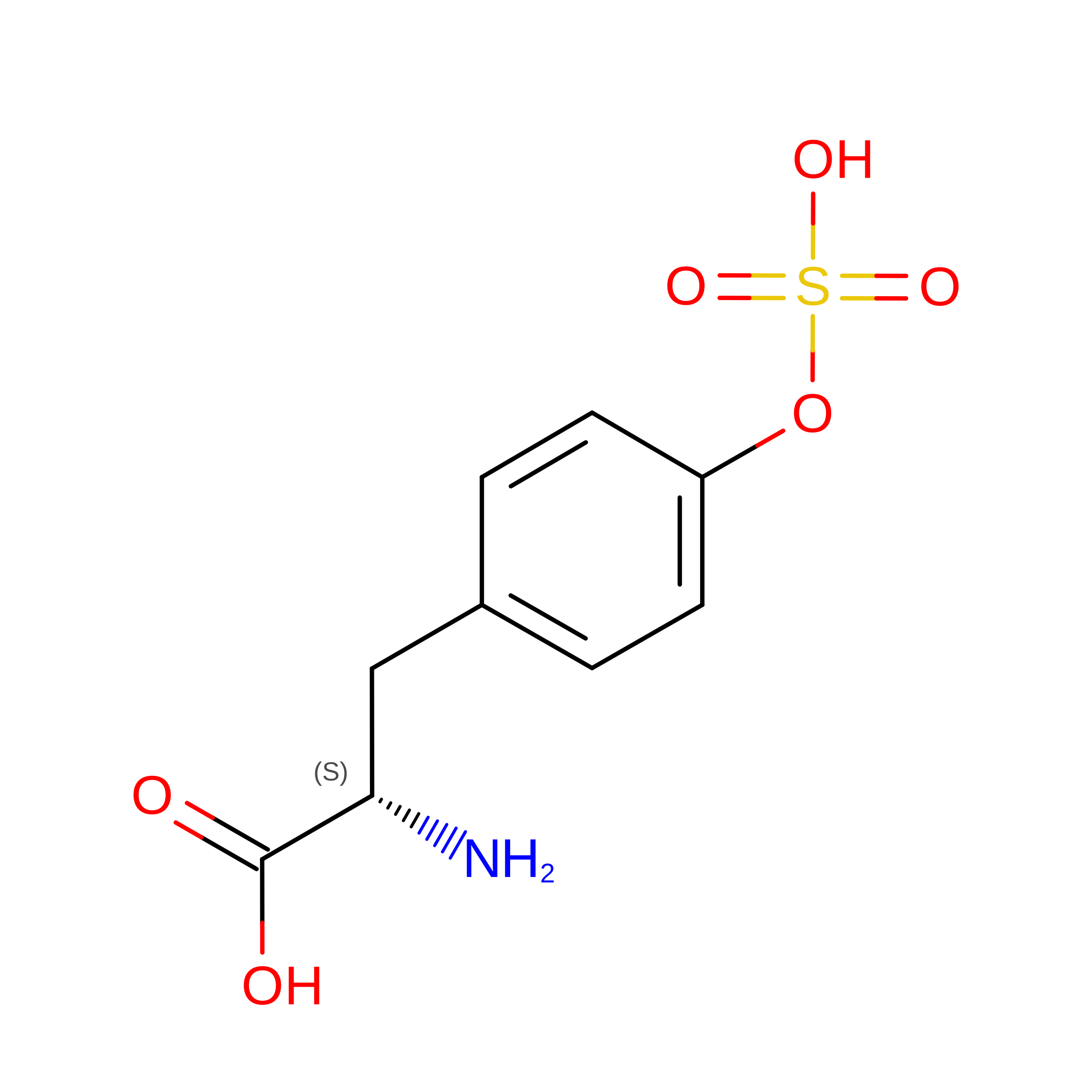
TYS
Query on TYS
G [auth H]L-PEPTIDE LINKINGC9 H11 N O6 STYR

Experimental Data & Validation

Experimental Data

  • Method: ELECTRON MICROSCOPY
  • Resolution: 3.48 Å
  • Aggregation State: PARTICLE 
  • Reconstruction Method: SINGLE PARTICLE 
EM Software:
TaskSoftware PackageVersion
MODEL REFINEMENTPHENIX

Structure Validation

View Full Validation Report


Entry History 

& Funding Information

Deposition Data


Funding OrganizationLocationGrant Number
National Institutes of Health/National Institute Of Allergy and Infectious Diseases (NIH/NIAID)United States--

Revision History  (Full details and data files)

  • Version 1.0: 2025-07-02
    Type: Initial release
  • Version 1.1: 2025-10-01
    Changes: Data collection, Database references