Comparative studies of seafood and reptile alpha- and beta-parvalbumins.
O'Malley, A., Ray, J.M., Kitlas, P., Ruethers, T., Kapingidza, A.B., Cierpicki, T., Lopata, A., Kowal, K., Chruszcz, M.(2024) Protein Sci 33: e5226-e5226
- PubMed: 39584689 Search on PubMedSearch on PubMed Central
- DOI: https://doi.org/10.1002/pro.5226
- Primary Citation Related Structures:
9B26, 9BAR, 9BB8, 9BCF - PubMed Abstract:
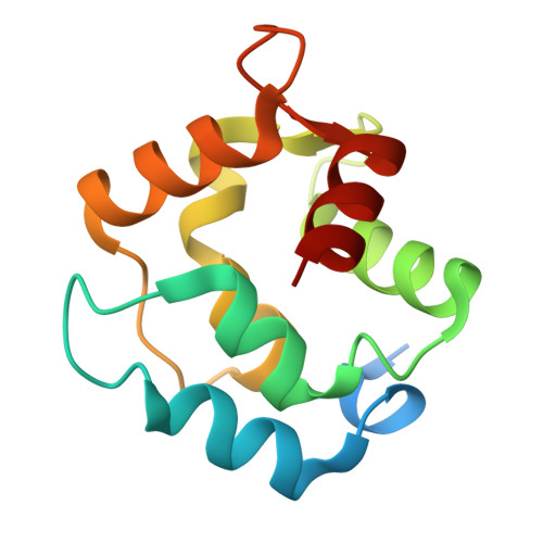
Small calcium-binding proteins such as parvalbumins (PVs) are major seafood and fish allergens. However, the impact of structural changes on their capacity to bind IgE has not been studied in detail. Therefore, fish and reptilian PVs, as well as human α-PV, were selected for biochemical, structural, and IgE binding studies. Likely due to their high solubility, crystallization proved difficult, so additional techniques were used to promote crystallization of the proteins. Novel crystal structures were determined for human PV, cod allergen Gad m 1.0201, saltwater crocodile allergen Cro p 1.0101, and the α-PV from thornback ray. β-PVs are considered the major fish allergens, while α-PVs are rarely categorized as allergens. To explain these differences, the results of structural and IgE binding studies were combined. This approach allowed us to provide new insight into IgE binding epitopes present on PVs, focusing on cross-reactivity among the selected α- and β-PVs. In addition, we have shown that these proteins display remarkable thermal stability across a range of pH conditions, which is relevant in the case of food allergens and food processing. Moreover, it is shown that the presence of calcium cations is critical for stability of the studied PVs via their protein folding, which has an impact on the formation of IgE binding epitopes. These studies shows the stability of fish and reptile PV allergens, and it allows for further evaluation of their IgE cross-reactivity.
- Department of Biochemistry and Molecular Biology, Michigan State University, East Lansing, Michigan, USA.
Organizational Affiliation: