9BAN | pdb_00009ban

The Anti-Mullerian Hormone prodomain in complex with the growth factor and 6E11 Fab in C1 symmetry


Experimental Data Snapshot

  • Method: ELECTRON MICROSCOPY
  • Resolution: 3.39 Å
  • Aggregation State: PARTICLE 
  • Reconstruction Method: SINGLE PARTICLE 

Starting Model: in silico
View more details

wwPDB Validation 3D Report Full Report

Validation slider image for 9BAN

This is version 1.0 of the entry. See complete history

Literature

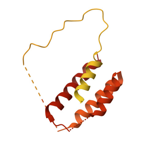
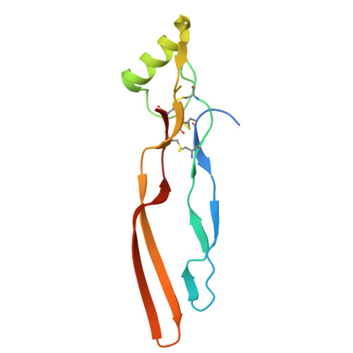
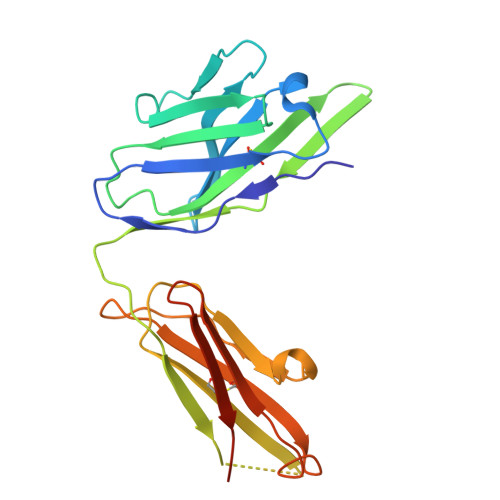
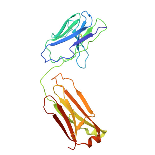
A divergent two-domain structure of the anti-Mullerian hormone prodomain.

Howard, J.A.Hok, L.Cate, R.L.Sanford, N.J.Hart, K.N.Leach, E.A.E.Bruening, A.S.Nagykery, N.Donahoe, P.K.Pepin, D.Thompson, T.B.

(2025) Proc Natl Acad Sci U S A 122: e2418088122-e2418088122

  • DOI: https://doi.org/10.1073/pnas.2418088122
  • Primary Citation Related Structures: 
    9BAN, 9BAO

  • PubMed Abstract: 

    TGFβ family ligands are synthesized as precursors consisting of an N-terminal prodomain and C-terminal growth factor (GF) signaling domain. After proteolytic processing, the prodomain typically remains noncovalently associated with the GF, sometimes forming a high-affinity latent procomplex that requires activation. For the TGFβ family ligand anti-Müllerian hormone (AMH), the prodomain maintains a high-affinity interaction with its GF that does not render it latent. While the prodomain can be displaced by the type II receptor, AMHR2, the nature of the GF:prodomain interaction and the mechanism of prodomain displacement by AMHR2 are currently unknown. We show here that the AMH prodomain exhibits an atypical two-domain structure, containing a dimerizing and a GF-binding domain connected through a flexible linker. Cryo-EM and genomic analyses show that the distinctive GF-binding domain, the result of an exon insertion 450 Mya, comprises a helical bundle and a belt-like structure which interact with the GF at the type II and I receptor binding sites, respectively. The dimerizing domain, which adopts a TGFβ-like propeptide fold, covalently connects two prodomains through intermolecular disulfide bonds. Disease mutations map to both the GF-binding and dimerization domains. Our results support a model where AMHR2 displaces the helical bundle and induces a conformational change in the GF, followed by release of the prodomain and engagement of the type I receptor. Collectively, this study shows that the AMH prodomain has evolved an atypical binding interaction with the GF that favors, without disrupting signaling, the maintenance of a noncovalent complex until receptors are engaged.


  • Organizational Affiliation
    • Department of Pharmacology, Physiology, and Neurobiology, University of Cincinnati, Cincinnati, OH 45267.

Macromolecule Content 

  • Total Structure Weight: 209.11 kDa 
  • Atom Count: 8,821 
  • Modeled Residue Count: 1,162 
  • Deposited Residue Count: 1,952 
  • Unique protein chains: 4

Macromolecules

Find similar proteins by:|  3D Structure
Entity ID: 1
MoleculeChains  Sequence LengthOrganismDetailsImage
Muellerian-inhibiting factor
A, C
427Homo sapiensMutation(s): 1 
Gene Names: AMHMIF
UniProt & NIH Common Fund Data Resources
Find proteins for P03971 (Homo sapiens)
Explore P03971 
Go to UniProtKB:  P03971
PHAROS:  P03971
GTEx:  ENSG00000104899 
Entity Groups
Sequence Clusters30% Identity50% Identity70% Identity90% Identity95% Identity100% Identity
UniProt GroupP03971
Sequence Annotations
Expand
Reference Sequence
Find similar proteins by:|  3D Structure
Entity ID: 2
MoleculeChains  Sequence LengthOrganismDetailsImage
Muellerian-inhibiting factor
B, D
109Homo sapiensMutation(s): 1 
Gene Names: AMHMIF
UniProt & NIH Common Fund Data Resources
Find proteins for P03971 (Homo sapiens)
Explore P03971 
Go to UniProtKB:  P03971
PHAROS:  P03971
GTEx:  ENSG00000104899 
Entity Groups
Sequence Clusters30% Identity50% Identity70% Identity90% Identity95% Identity100% Identity
UniProt GroupP03971
Sequence Annotations
Expand
Reference Sequence
Find similar proteins by:|  3D Structure
Entity ID: 3
MoleculeChains  Sequence LengthOrganismDetailsImage
6E11 Antibody IgG2A Heavy Chain
E, G
227Mus musculusMutation(s): 0 
Find similar proteins by:|  3D Structure
Entity ID: 4
MoleculeChains  Sequence LengthOrganismDetailsImage
6E11 Antibody kappa Light Chain
F, H
213Mus musculusMutation(s): 0 

Experimental Data & Validation

Experimental Data

  • Method: ELECTRON MICROSCOPY
  • Resolution: 3.39 Å
  • Aggregation State: PARTICLE 
  • Reconstruction Method: SINGLE PARTICLE 
EM Software:
TaskSoftware PackageVersion
MODEL REFINEMENTPHENIX
RECONSTRUCTIONcryoSPARC

Structure Validation

View Full Validation Report


Entry History 

& Funding Information

Deposition Data


Funding OrganizationLocationGrant Number
National Institutes of Health/Eunice Kennedy Shriver National Institute of Child Health & Human Development (NIH/NICHD)United StatesHD105818

Revision History  (Full details and data files)

  • Version 1.0: 2025-01-29
    Type: Initial release