9B4H

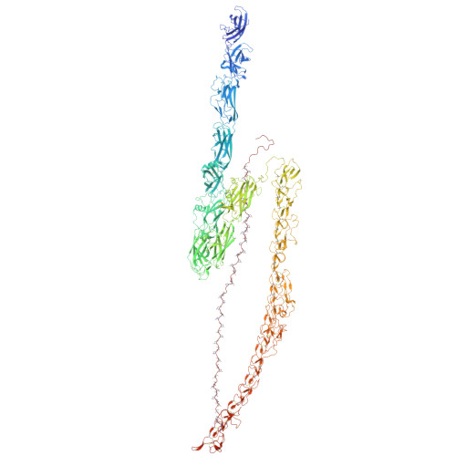
Chlamydomonas reinhardtii mastigoneme filament

  • Classification: STRUCTURAL PROTEIN
  • Organism(s): Chlamydomonas reinhardtii
  • Mutation(s): No 

  • Deposited: 2024-03-20 Released: 2024-04-10 
  • Deposition Author(s): Dai, J., Ma, M., Zhang, R., Brown, A.
  • Funding Organization(s): National Institutes of Health/National Institute of General Medical Sciences (NIH/NIGMS), National Institutes of Health/National Heart, Lung, and Blood Institute (NIH/NHLBI)

Experimental Data Snapshot

  • Method: ELECTRON MICROSCOPY
  • Resolution: 3.10 Å
  • Aggregation State: FILAMENT 
  • Reconstruction Method: SINGLE PARTICLE 

Starting Model: in silico
View more details

wwPDB Validation   3D Report Full Report


This is version 1.0 of the entry. See complete history


Literature

Mastigoneme structure reveals insights into the O-linked glycosylation code of native hydroxyproline-rich helices.

Dai, J.Ma, M.Niu, Q.Eisert, R.J.Wang, X.Das, P.Lechtreck, K.F.Dutcher, S.K.Zhang, R.Brown, A.

(2024) Cell 

  • DOI: https://doi.org/10.1016/j.cell.2024.03.005
  • Primary Citation of Related Structures:  
    9B4H

  • PubMed Abstract: 

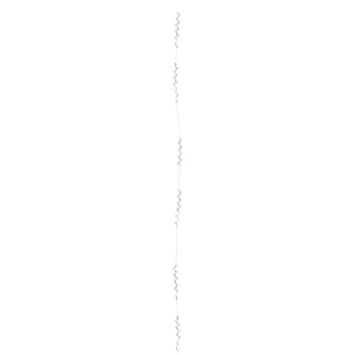
    Hydroxyproline-rich glycoproteins (HRGPs) are a ubiquitous class of protein in the extracellular matrices and cell walls of plants and algae, yet little is known of their native structures or interactions. Here, we used electron cryomicroscopy (cryo-EM) to determine the structure of the hydroxyproline-rich mastigoneme, an extracellular filament isolated from the cilia of the alga Chlamydomonas reinhardtii. The structure demonstrates that mastigonemes are formed from two HRGPs (a filament of MST1 wrapped around a single copy of MST3) that both have hyperglycosylated poly(hydroxyproline) helices. Within the helices, O-linked glycosylation of the hydroxyproline residues and O-galactosylation of interspersed serine residues create a carbohydrate casing. Analysis of the associated glycans reveals how the pattern of hydroxyproline repetition determines the type and extent of glycosylation. MST3 possesses a PKD2-like transmembrane domain that forms a heteromeric polycystin-like cation channel with PKD2 and SIP, explaining how mastigonemes are tethered to ciliary membranes.


  • Organizational Affiliation

    Department of Biological Chemistry and Molecular Pharmacology, Blavatnik Institute, Harvard Medical School, Boston, MA, USA.


Macromolecules
Find similar proteins by:  (by identity cutoff)  |  3D Structure
Entity ID: 1
MoleculeChains Sequence LengthOrganismDetailsImage
Tyrosine-protein kinase ephrin type A/B receptor-like domain-containing proteinA,
C [auth B]
1,987Chlamydomonas reinhardtiiMutation(s): 0 
UniProt
Find proteins for A8J9H7 (Chlamydomonas reinhardtii)
Explore A8J9H7 
Go to UniProtKB:  A8J9H7
Entity Groups  
Sequence Clusters30% Identity50% Identity70% Identity90% Identity95% Identity100% Identity
UniProt GroupA8J9H7
Glycosylation
Glycosylation Sites: 16
Sequence Annotations
Expand
  • Reference Sequence
Find similar proteins by:  (by identity cutoff)  |  3D Structure
Entity ID: 2
MoleculeChains Sequence LengthOrganismDetailsImage
C-type lectin domain-containing proteinB [auth X]8,572Chlamydomonas reinhardtiiMutation(s): 0 
UniProt
Find proteins for A0A2K3DRP3 (Chlamydomonas reinhardtii)
Explore A0A2K3DRP3 
Go to UniProtKB:  A0A2K3DRP3
Entity Groups  
Sequence Clusters30% Identity50% Identity70% Identity90% Identity95% Identity100% Identity
UniProt GroupA0A2K3DRP3
Sequence Annotations
Expand
  • Reference Sequence
Oligosaccharides

Help

Entity ID: 3
MoleculeChains Length2D Diagram Glycosylation3D Interactions
2-acetamido-2-deoxy-beta-D-glucopyranose-(1-4)-2-acetamido-2-deoxy-beta-D-glucopyranose
AB [auth a2],
BB [auth a3],
D [auth A1],
E [auth A2],
F [auth A3],
AB [auth a2],
BB [auth a3],
D [auth A1],
E [auth A2],
F [auth A3],
ZA [auth a1]
2N/AN-Glycosylation
Entity ID: 4
MoleculeChains Length2D Diagram Glycosylation3D Interactions
beta-L-arabinofuranose-(1-2)-beta-L-arabinofuranose
AA [auth C7],
AC [auth d2],
AD [auth d0],
BA [auth C8],
BC [auth d3],
AA [auth C7],
AC [auth d2],
AD [auth d0],
BA [auth C8],
BC [auth d3],
BD [auth F5],
CA [auth C9],
CC [auth d4],
CD [auth F2],
DA [auth D0],
DB [auth a5],
DC [auth d5],
DD [auth E7],
EA [auth D1],
EC [auth e0],
FA [auth D2],
FB [auth a7],
FC [auth e1],
G [auth A4],
GA [auth D3],
GC [auth e2],
H [auth A5],
HA [auth D4],
HB [auth b0],
HC [auth e4],
IA [auth D5],
IB [auth b1],
IC [auth e5],
J [auth A7],
JA [auth D6],
JB [auth b3],
JC [auth e8],
KA [auth D7],
KB [auth b4],
L [auth B0],
LA [auth D8],
LB [auth b5],
LC [auth f0],
M [auth B1],
MA [auth D9],
MC [auth f3],
N [auth B3],
NA [auth E0],
NB [auth b7],
NC [auth f5],
O [auth B4],
OA [auth E1],
OB [auth b9],
P [auth B5],
PA [auth E2],
PB [auth c0],
QA [auth E4],
QB [auth c1],
QC [auth f8],
R [auth B7],
RA [auth E5],
RB [auth c2],
RC [auth E3],
S [auth B9],
SB [auth c3],
SC [auth f2],
T [auth C0],
TA [auth E8],
TB [auth c4],
TC [auth e7],
U [auth C1],
UA [auth F0],
UB [auth c5],
UC [auth e6],
V [auth C2],
VA [auth F3],
VB [auth c6],
VC [auth e3],
W [auth C3],
WB [auth c7],
WC [auth d9],
X [auth C4],
XB [auth c8],
XC [auth d7],
Y [auth C5],
YA [auth F8],
YB [auth c9],
YC [auth d8],
Z [auth C6],
ZB [auth d1],
ZC [auth d6]
2N/AN/A
Entity ID: 5
MoleculeChains Length2D Diagram Glycosylation3D Interactions
beta-L-arabinofuranose-(1-2)-beta-L-arabinofuranose-(5-5)-beta-L-arabinofuranose-(1-2)-beta-L-arabinofuranose
EB [auth a6],
GB [auth a9],
I [auth A6],
K [auth A9],
MB [auth b6],
EB [auth a6],
GB [auth a9],
I [auth A6],
K [auth A9],
MB [auth b6],
OC [auth f6],
PC [auth f7],
Q [auth B6],
WA [auth F6],
XA [auth F7]
4N/AN/A
Entity ID: 6
MoleculeChains Length2D Diagram Glycosylation3D Interactions
beta-L-arabinofuranose-(1-2)-beta-L-arabinofuranose-(5-5)-beta-L-arabinofuranose-(1-2)-beta-L-arabinofuranose-(5-5)-beta-L-arabinofuranose-(1-2)-beta-L-arabinofuranose-(5-5)-beta-L-arabinofuranose-(1-2)-beta-L-arabinofuranoseSA [auth E6]8N/AN/A
Entity ID: 7
MoleculeChains Length2D Diagram Glycosylation3D Interactions
beta-L-arabinofuranose-(1-4)-beta-L-ribulofuranoseCB [auth a4]2N/AN/A
Entity ID: 8
MoleculeChains Length2D Diagram Glycosylation3D Interactions
beta-L-arabinofuranose-(1-2)-beta-L-arabinofuranose-(5-5)-beta-L-arabinofuranose-(1-2)-beta-L-arabinofuranose-(5-5)-beta-L-arabinofuranose-(1-2)-beta-L-arabinofuranoseKC [auth e9]6N/AN/A
Small Molecules
Ligands 2 Unique
IDChains Name / Formula / InChI Key2D Diagram3D Interactions
GLA (Subject of Investigation/LOI)
Query on GLA

Download Ideal Coordinates CCD File 
BI [auth B]
CI [auth B]
DI [auth B]
EI [auth B]
FI [auth B]
BI [auth B],
CI [auth B],
DI [auth B],
EI [auth B],
FI [auth B],
GI [auth B],
HI [auth B],
II [auth B],
JF [auth A],
JI [auth B],
KF [auth A],
KI [auth B],
LF [auth A],
LI [auth B],
MF [auth A],
MI [auth B],
NF [auth A],
NI [auth B],
OF [auth A],
PF [auth A],
QF [auth A],
RF [auth A],
SF [auth A],
TF [auth A],
UF [auth A],
VF [auth A]
alpha-D-galactopyranose
C6 H12 O6
WQZGKKKJIJFFOK-PHYPRBDBSA-N
A1AIO (Subject of Investigation/LOI)
Query on A1AIO

Download Ideal Coordinates CCD File 
AE [auth A]
AF [auth A]
AG [auth B]
AH [auth B]
AI [auth B]
AE [auth A],
AF [auth A],
AG [auth B],
AH [auth B],
AI [auth B],
BE [auth A],
BF [auth A],
BG [auth B],
BH [auth B],
CE [auth A],
CF [auth A],
CG [auth B],
CH [auth B],
DE [auth A],
DF [auth A],
DG [auth B],
DH [auth B],
ED [auth A],
EE [auth A],
EF [auth A],
EG [auth B],
EH [auth B],
FD [auth A],
FE [auth A],
FF [auth A],
FG [auth B],
FH [auth B],
GD [auth A],
GE [auth A],
GF [auth A],
GG [auth B],
GH [auth B],
HD [auth A],
HE [auth A],
HF [auth A],
HG [auth B],
HH [auth B],
ID [auth A],
IE [auth A],
IF [auth A],
IG [auth B],
IH [auth B],
JD [auth A],
JE [auth A],
JG [auth B],
JH [auth B],
KD [auth A],
KE [auth A],
KG [auth B],
KH [auth B],
LD [auth A],
LE [auth A],
LG [auth B],
LH [auth B],
MD [auth A],
ME [auth A],
MG [auth B],
MH [auth B],
ND [auth A],
NE [auth A],
NG [auth B],
NH [auth B],
OD [auth A],
OE [auth A],
OG [auth B],
OH [auth B],
PD [auth A],
PE [auth A],
PG [auth B],
PH [auth B],
QD [auth A],
QE [auth A],
QG [auth B],
QH [auth B],
RD [auth A],
RE [auth A],
RG [auth B],
RH [auth B],
SD [auth A],
SE [auth A],
SG [auth B],
SH [auth B],
TD [auth A],
TE [auth A],
TG [auth B],
TH [auth B],
UD [auth A],
UE [auth A],
UG [auth B],
UH [auth B],
VD [auth A],
VE [auth A],
VG [auth B],
VH [auth B],
WD [auth A],
WE [auth A],
WF [auth B],
WG [auth B],
WH [auth B],
XD [auth A],
XE [auth A],
XF [auth B],
XG [auth B],
XH [auth B],
YD [auth A],
YE [auth A],
YF [auth B],
YG [auth B],
YH [auth B],
ZD [auth A],
ZE [auth A],
ZF [auth B],
ZG [auth B],
ZH [auth B]
beta-L-glucofuranose
C6 H12 O6
AVVWPBAENSWJCB-RUTHBDMASA-N
Modified Residues  1 Unique
IDChains TypeFormula2D DiagramParent
HYP
Query on HYP
A,
C [auth B]
L-PEPTIDE LINKINGC5 H9 N O3PRO
Experimental Data & Validation

Experimental Data

  • Method: ELECTRON MICROSCOPY
  • Resolution: 3.10 Å
  • Aggregation State: FILAMENT 
  • Reconstruction Method: SINGLE PARTICLE 
EM Software:
TaskSoftware PackageVersion
MODEL REFINEMENTPHENIX1.21_5207:

Structure Validation

View Full Validation Report


Entry History & Funding Information

Deposition Data


Funding OrganizationLocationGrant Number
National Institutes of Health/National Institute of General Medical Sciences (NIH/NIGMS)United StatesR01GM141109
National Institutes of Health/National Institute of General Medical Sciences (NIH/NIGMS)United StatesR01GM143183
National Institutes of Health/National Institute of General Medical Sciences (NIH/NIGMS)United StatesR01GM138854
National Institutes of Health/National Heart, Lung, and Blood Institute (NIH/NHLBI)United StatesR01HL128370
National Institutes of Health/National Institute of General Medical Sciences (NIH/NIGMS)United StatesR35GM131909
National Institutes of Health/National Institute of General Medical Sciences (NIH/NIGMS)United StatesR01GM139856
National Institutes of Health/National Institute of General Medical Sciences (NIH/NIGMS)United StatesR35GM152057

Revision History  (Full details and data files)

  • Version 1.0: 2024-04-10
    Type: Initial release