8VQ3

CDK2-CyclinE1 in complex with allosteric inhibitor I-198.


Experimental Data Snapshot

  • Method: X-RAY DIFFRACTION
  • Resolution: 1.84 Å
  • R-Value Free: 0.217 
  • R-Value Work: 0.199 
  • R-Value Observed: 0.200 

Starting Model: experimental
View more details

wwPDB Validation   3D Report Full Report


Ligand Structure Quality Assessment 


This is version 1.2 of the entry. See complete history


Literature

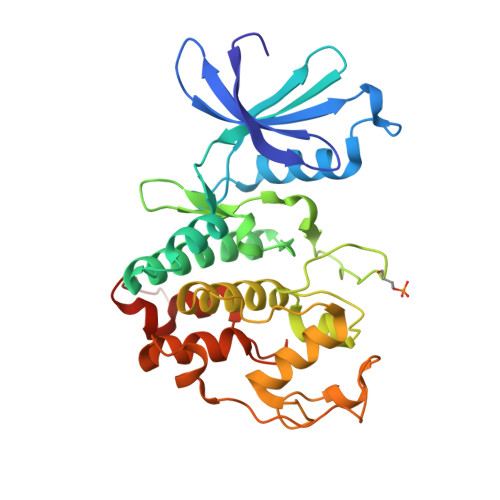
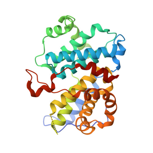
An allosteric cyclin E-CDK2 site mapped by paralog hopping with covalent probes.

Zhang, Y.Liu, Z.Hirschi, M.Brodsky, O.Johnson, E.Won, S.J.Nagata, A.Bezwada, D.Petroski, M.D.Majmudar, J.D.Niessen, S.VanArsdale, T.Gilbert, A.M.Hayward, M.M.Stewart, A.E.Nager, A.R.Melillo, B.Cravatt, B.F.

(2024) Nat Chem Biol 

  • DOI: https://doi.org/10.1038/s41589-024-01738-7
  • Primary Citation of Related Structures:  
    8VQ3, 8VQ4

  • PubMed Abstract: 

    More than half of the ~20,000 protein-encoding human genes have paralogs. Chemical proteomics has uncovered many electrophile-sensitive cysteines that are exclusive to subsets of paralogous proteins. Here we explore whether such covalent compound-cysteine interactions can be used to discover ligandable pockets in paralogs lacking the cysteine. Leveraging the covalent ligandability of C109 in the cyclin CCNE2, we substituted the corresponding residue in paralog CCNE1 to cysteine (N112C) and found through activity-based protein profiling that this mutant reacts stereoselectively and site-specifically with tryptoline acrylamides. We then converted the tryptoline acrylamide-CCNE1-N112C interaction into in vitro NanoBRET (bioluminescence resonance energy transfer) and in cellulo activity-based protein profiling assays capable of identifying compounds that reversibly inhibit both the N112C mutant and wild-type CCNE1:CDK2 (cyclin-dependent kinase 2) complexes. X-ray crystallography revealed a cryptic allosteric pocket at the CCNE1:CDK2 interface adjacent to N112 that binds the reversible inhibitors. Our findings, thus, show how electrophile-cysteine interactions mapped by chemical proteomics can extend the understanding of protein ligandability beyond covalent chemistry.


  • Organizational Affiliation

    Department of Chemistry, The Scripps Research Institute, La Jolla, CA, USA.


Macromolecules
Find similar proteins by:  (by identity cutoff)  |  3D Structure
Entity ID: 1
MoleculeChains Sequence LengthOrganismDetailsImage
Cyclin-dependent kinase 2298Homo sapiensMutation(s): 0 
Gene Names: CDK2CDKN2
EC: 2.7.11.22
UniProt & NIH Common Fund Data Resources
Find proteins for P24941 (Homo sapiens)
Explore P24941 
Go to UniProtKB:  P24941
PHAROS:  P24941
GTEx:  ENSG00000123374 
Entity Groups  
Sequence Clusters30% Identity50% Identity70% Identity90% Identity95% Identity100% Identity
UniProt GroupP24941
Sequence Annotations
Expand
  • Reference Sequence
Find similar proteins by:  (by identity cutoff)  |  3D Structure
Entity ID: 2
MoleculeChains Sequence LengthOrganismDetailsImage
G1/S-specific cyclin-E1285Homo sapiensMutation(s): 0 
Gene Names: CCNE1CCNE
UniProt & NIH Common Fund Data Resources
Find proteins for P24864 (Homo sapiens)
Explore P24864 
Go to UniProtKB:  P24864
PHAROS:  P24864
GTEx:  ENSG00000105173 
Entity Groups  
Sequence Clusters30% Identity50% Identity70% Identity90% Identity95% Identity100% Identity
UniProt GroupP24864
Sequence Annotations
Expand
  • Reference Sequence
Small Molecules
Ligands 1 Unique
IDChains Name / Formula / InChI Key2D Diagram3D Interactions
A1AC4
Query on A1AC4

Download Ideal Coordinates CCD File 
C [auth A](8R)-N-[(2S,3R)-3-(cyclohexylmethoxy)-1-(morpholin-4-yl)-1-oxobutan-2-yl]-2-[(1S)-2,2-dimethylcyclopropane-1-carbonyl]-6-(1,3-thiazole-5-carbonyl)-2,6-diazaspiro[3.4]octane-8-carboxamide
C32 H47 N5 O6 S
NGKYDOYICDCFRF-NZMKUBJDSA-N
Modified Residues  1 Unique
IDChains TypeFormula2D DiagramParent
TPO
Query on TPO
A
L-PEPTIDE LINKINGC4 H10 N O6 PTHR
Experimental Data & Validation

Experimental Data

  • Method: X-RAY DIFFRACTION
  • Resolution: 1.84 Å
  • R-Value Free: 0.217 
  • R-Value Work: 0.199 
  • R-Value Observed: 0.200 
  • Space Group: P 41 21 2
Unit Cell:
Length ( Å )Angle ( ˚ )
a = 101.388α = 90
b = 101.388β = 90
c = 151.613γ = 90
Software Package:
Software NamePurpose
PHENIXrefinement
XDSdata reduction
Aimlessdata scaling
PHASERphasing

Structure Validation

View Full Validation Report


Ligand Structure Quality Assessment 


Entry History & Funding Information

Deposition Data


Funding OrganizationLocationGrant Number
National Institutes of Health/National Cancer Institute (NIH/NCI)United StatesCA231991
PfizerUnited States--

Revision History  (Full details and data files)

  • Version 1.0: 2024-01-31
    Type: Initial release
  • Version 1.1: 2024-02-14
    Changes: Database references
  • Version 1.2: 2024-10-16
    Changes: Database references, Structure summary