8VCR | pdb_00008vcr

SARS-CoV-2 Spike S2 bound to Fab 54043-5


Experimental Data Snapshot

  • Method: ELECTRON MICROSCOPY
  • Resolution: 3.00 Å
  • Aggregation State: PARTICLE 
  • Reconstruction Method: SINGLE PARTICLE 

wwPDB Validation 3D Report Full Report

Validation slider image for 8VCR

This is version 1.3 of the entry. See complete history

Literature

Discovery and characterization of a pan-betacoronavirus S2-binding antibody.

Johnson, N.V.Wall, S.C.Kramer, K.J.Holt, C.M.Periasamy, S.Richardson, S.I.Manamela, N.P.Suryadevara, N.Andreano, E.Paciello, I.Pierleoni, G.Piccini, G.Huang, Y.Ge, P.Allen, J.D.Uno, N.Shiakolas, A.R.Pilewski, K.A.Nargi, R.S.Sutton, R.E.Abu-Shmais, A.A.Parks, R.Haynes, B.F.Carnahan, R.H.Crowe Jr., J.E.Montomoli, E.Rappuoli, R.Bukreyev, A.Ross, T.M.Sautto, G.A.McLellan, J.S.Georgiev, I.S.

(2024) Structure 32: 1893

  • DOI: https://doi.org/10.1016/j.str.2024.08.022
  • Primary Citation Related Structures: 
    8VCR

  • PubMed Abstract: 

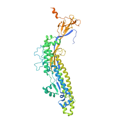
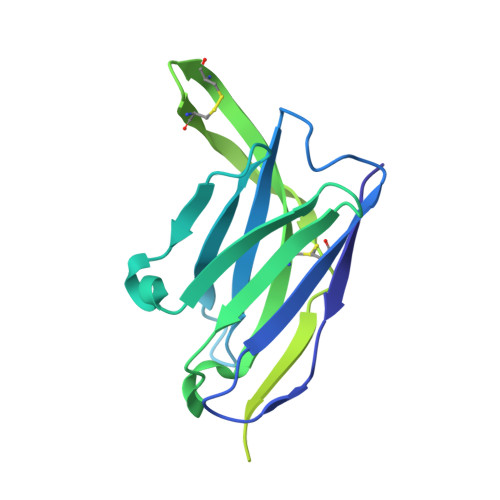
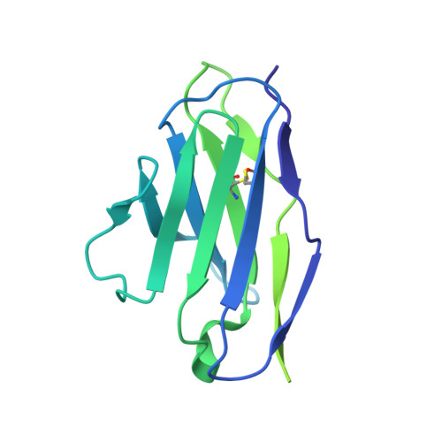
    The continued emergence of deadly human coronaviruses from animal reservoirs highlights the need for pan-coronavirus interventions for effective pandemic preparedness. Here, using linking B cell receptor to antigen specificity through sequencing (LIBRA-seq), we report a panel of 50 coronavirus antibodies isolated from human B cells. Of these, 54043-5 was shown to bind the S2 subunit of spike proteins from alpha-, beta-, and deltacoronaviruses. A cryoelectron microscopy (cryo-EM) structure of 54043-5 bound to the prefusion S2 subunit of the severe acute respiratory syndrome coronavirus 2 (SARS-CoV-2) spike defined an epitope at the apex of S2 that is highly conserved among betacoronaviruses. Although non-neutralizing, 54043-5 induced Fc-dependent antiviral responses in vitro, including antibody-dependent cellular cytotoxicity (ADCC) and antibody-dependent cellular phagocytosis (ADCP). In murine SARS-CoV-2 challenge studies, protection against disease was observed after introduction of Leu234Ala, Leu235Ala, and Pro329Gly (LALA-PG) substitutions in the Fc region of 54043-5. Together, these data provide new insights into the protective mechanisms of non-neutralizing antibodies and define a broadly conserved epitope within the S2 subunit.


  • Organizational Affiliation
    • Department of Molecular Biosciences, The University of Texas at Austin, Austin, TX 78712, USA.

Macromolecule Content 

  • Total Structure Weight: 347.71 kDa 
  • Atom Count: 15,987 
  • Modeled Residue Count: 2,040 
  • Deposited Residue Count: 3,132 
  • Unique protein chains: 3

Macromolecules

Find similar proteins by:|  3D Structure
Entity ID: 1
MoleculeChains  Sequence LengthOrganismDetailsImage
Spike protein S2A,
H [auth B],
I [auth C]
597Severe acute respiratory syndrome coronavirus 2Mutation(s): 9 
Gene Names: S2
UniProt
Find proteins for P0DTC2 (Severe acute respiratory syndrome coronavirus 2)
Explore P0DTC2 
Go to UniProtKB:  P0DTC2
Entity Groups
Sequence Clusters30% Identity50% Identity70% Identity90% Identity95% Identity100% Identity
UniProt GroupP0DTC2
Glycosylation
Glycosylation Sites: 5Go to GlyGen: P0DTC2-1
Sequence Annotations
Expand
Reference Sequence
Find similar proteins by:|  3D Structure
Entity ID: 2
MoleculeChains  Sequence LengthOrganismDetailsImage
54043-5 Fab Heavy ChainB [auth D],
C [auth E],
F [auth H]
233Homo sapiensMutation(s): 0 
Find similar proteins by:|  3D Structure
Entity ID: 3
MoleculeChains  Sequence LengthOrganismDetailsImage
54043-5 Fab Light ChainD [auth F],
E [auth G],
G [auth L]
214Homo sapiensMutation(s): 0 

Oligosaccharides

Help  
Entity ID: 4
MoleculeChains Length2D Diagram GlycosylationD Interactions
2-acetamido-2-deoxy-beta-D-glucopyranose-(1-4)-2-acetamido-2-deoxy-beta-D-glucopyranoseJ [auth I],
K [auth J],
L [auth K]
2N-Glycosylation
Glycosylation Resources
GlyTouCan: G42666HT
GlyCosmos: G42666HT
GlyGen: G42666HT

Small Molecules

Ligands 1 Unique
IDChains Name / Formula / InChI Key2D Diagram3D Interactions
NAG

Query on NAG


Download:Ideal Coordinates CCD File
AA [auth C]
M [auth A]
N [auth A]
O [auth A]
P [auth A]
AA [auth C],
M [auth A],
N [auth A],
O [auth A],
P [auth A],
Q [auth D],
R [auth E],
S [auth H],
T [auth B],
U [auth B],
V [auth B],
W [auth B],
X [auth C],
Y [auth C],
Z [auth C]
2-acetamido-2-deoxy-beta-D-glucopyranose
C8 H15 N O6
OVRNDRQMDRJTHS-FMDGEEDCSA-N

Experimental Data & Validation

Experimental Data

  • Method: ELECTRON MICROSCOPY
  • Resolution: 3.00 Å
  • Aggregation State: PARTICLE 
  • Reconstruction Method: SINGLE PARTICLE 
EM Software:
TaskSoftware PackageVersion
MODEL REFINEMENTPHENIX1.18.2_3874:

Structure Validation

View Full Validation Report


Entry History 

& Funding Information

Deposition Data


Funding OrganizationLocationGrant Number
Not funded--

Revision History  (Full details and data files)

  • Version 1.0: 2024-07-31
    Type: Initial release
  • Version 1.1: 2024-10-16
    Changes: Data collection, Database references, Structure summary
  • Version 1.2: 2024-10-23
    Changes: Data collection
  • Version 1.3: 2024-11-27
    Changes: Data collection, Database references