8TYS

Adaptive mechanism of collagen IV scaffold assembly in Drosophila: crystal structure of tissue-extracted NC1 hexamer

  • Classification: STRUCTURAL PROTEIN
  • Organism(s): Drosophila melanogaster
  • Mutation(s): No 

  • Deposited: 2023-08-25 Released: 2023-11-08 
  • Deposition Author(s): Boudko, S.P.
  • Funding Organization(s): National Institutes of Health/National Institute of Diabetes and Digestive and Kidney Disease (NIH/NIDDK)

Experimental Data Snapshot

  • Method: X-RAY DIFFRACTION
  • Resolution: 2.90 Å
  • R-Value Free: 0.277 
  • R-Value Work: 0.241 
  • R-Value Observed: 0.243 

wwPDB Validation   3D Report Full Report


This is version 1.1 of the entry. See complete history


Literature

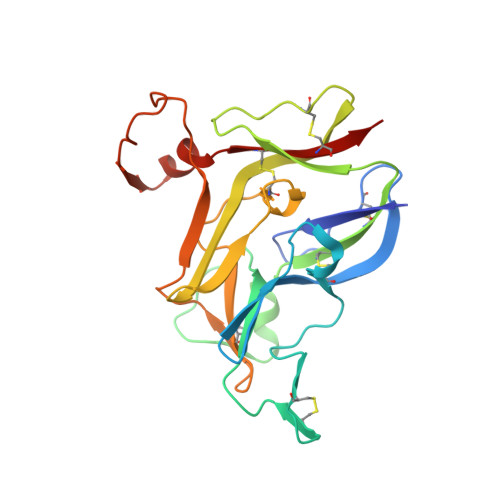
Collagen IV of basement membranes: IV. Adaptive mechanism of collagen IV scaffold assembly in Drosophila.

Summers, J.A.Yarbrough, M.Liu, M.McDonald, W.H.Hudson, B.G.Pastor-Pareja, J.C.Boudko, S.P.

(2023) J Biol Chem 299: 105394-105394

  • DOI: https://doi.org/10.1016/j.jbc.2023.105394
  • Primary Citation of Related Structures:  
    8TXN, 8TYS

  • PubMed Abstract: 

    Collagen IV is an essential structural protein in all metazoans. It provides a scaffold for the assembly of basement membranes, a specialized form of extracellular matrix, which anchors and signals cells and provides microscale tensile strength. Defective scaffolds cause basement membrane destabilization and tissue dysfunction. Scaffolds are composed of α-chains that coassemble into triple-helical protomers of distinct chain compositions, which in turn oligomerize into supramolecular scaffolds. Chloride ions mediate the oligomerization via NC1 trimeric domains, forming an NC1 hexamer at the protomer-protomer interface. The chloride concentration-"chloride pressure"-on the outside of cells is a primordial innovation that drives the assembly and dynamic stabilization of collagen IV scaffolds. However, a Cl-independent mechanism is operative in Ctenophora, Ecdysozoa, and Rotifera, which suggests evolutionary adaptations to environmental or tissue conditions. An understanding of these exceptions, such as the example of Drosophila, could shed light on the fundamentals of how NC1 trimers direct the oligomerization of protomers into scaffolds. Here, we investigated the NC1 assembly of Drosophila. We solved the crystal structure of the NC1 hexamer, determined the chain composition of protomers, and found that Drosophila adapted an evolutionarily unique mechanism of scaffold assembly that requires divalent cations. By studying the Drosophila case we highlighted the mechanistic role of chloride pressure for maintaining functionality of the NC1 domain in humans. Moreover, we discovered that the NC1 trimers encode information for homing protomers to distant tissue locations, providing clues for the development of protein replacement therapy for collagen IV genetic diseases.


  • Organizational Affiliation

    Aspirnaut Program, Vanderbilt University Medical Center, Nashville, Tennessee, USA.


Macromolecules
Find similar proteins by:  (by identity cutoff)  |  3D Structure
Entity ID: 1
MoleculeChains Sequence LengthOrganismDetailsImage
Collagen alpha-1(IV) chain
A, C, D, F
230Drosophila melanogasterMutation(s): 0 
UniProt
Find proteins for P08120 (Drosophila melanogaster)
Explore P08120 
Go to UniProtKB:  P08120
Entity Groups  
Sequence Clusters30% Identity50% Identity70% Identity90% Identity95% Identity100% Identity
UniProt GroupP08120
Sequence Annotations
Expand
  • Reference Sequence
Find similar proteins by:  (by identity cutoff)  |  3D Structure
Entity ID: 2
MoleculeChains Sequence LengthOrganismDetailsImage
Collagen IV, chain Viking
B, E
230Drosophila melanogasterMutation(s): 0 
UniProt
Find proteins for Q9VMV5 (Drosophila melanogaster)
Explore Q9VMV5 
Go to UniProtKB:  Q9VMV5
Entity Groups  
Sequence Clusters30% Identity50% Identity70% Identity90% Identity95% Identity100% Identity
UniProt GroupQ9VMV5
Sequence Annotations
Expand
  • Reference Sequence
Experimental Data & Validation

Experimental Data

  • Method: X-RAY DIFFRACTION
  • Resolution: 2.90 Å
  • R-Value Free: 0.277 
  • R-Value Work: 0.241 
  • R-Value Observed: 0.243 
  • Space Group: P 63
Unit Cell:
Length ( Å )Angle ( ˚ )
a = 145.209α = 90
b = 145.209β = 90
c = 106.491γ = 120
Software Package:
Software NamePurpose
PHENIXrefinement
iMOSFLMdata reduction
pointlessdata scaling
AMoREphasing

Structure Validation

View Full Validation Report


Entry History & Funding Information

Deposition Data

  • Released Date: 2023-11-08 
  • Deposition Author(s): Boudko, S.P.

Funding OrganizationLocationGrant Number
National Institutes of Health/National Institute of Diabetes and Digestive and Kidney Disease (NIH/NIDDK)United StatesR01DK018381
National Institutes of Health/National Institute of Diabetes and Digestive and Kidney Disease (NIH/NIDDK)United StatesR01DK131101

Revision History  (Full details and data files)

  • Version 1.0: 2023-11-08
    Type: Initial release
  • Version 1.1: 2023-12-06
    Changes: Database references