8TRR | pdb_00008trr

T cell recognition of citrullinated vimentin peptide presented by HLA-DR4


Experimental Data Snapshot

  • Method: X-RAY DIFFRACTION
  • Resolution: 2.65 Å
  • R-Value Free: 
    0.244 (Depositor), 0.237 (DCC) 
  • R-Value Work: 
    0.209 (Depositor) 
  • R-Value Observed: 
    0.210 (Depositor) 

Starting Model: experimental
View more details

wwPDB Validation 3D Report Full Report

Validation slider image for 8TRR

Ligand Structure Quality Assessment 


This is version 1.2 of the entry. See complete history

Literature

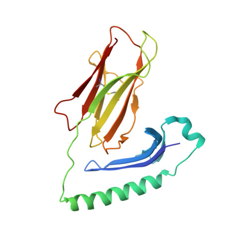
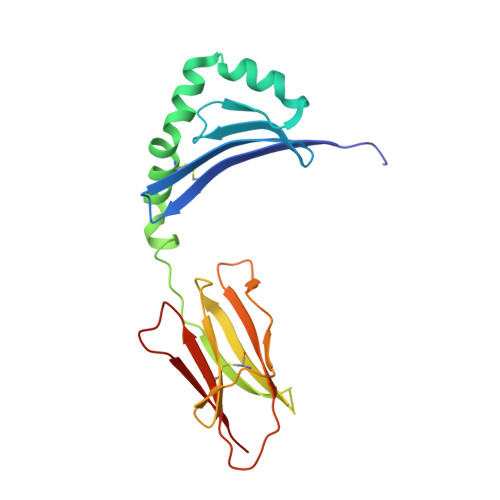
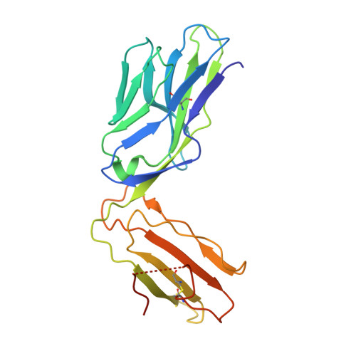
The molecular basis underlying T cell specificity towards citrullinated epitopes presented by HLA-DR4.

Loh, T.J.Lim, J.J.Jones, C.M.Dao, H.T.Tran, M.T.Baker, D.G.La Gruta, N.L.Reid, H.H.Rossjohn, J.

(2024) Nat Commun 15: 6201-6201

  • DOI: https://doi.org/10.1038/s41467-024-50511-w
  • Primary Citation Related Structures: 
    8TRL, 8TRQ, 8TRR

  • PubMed Abstract: 

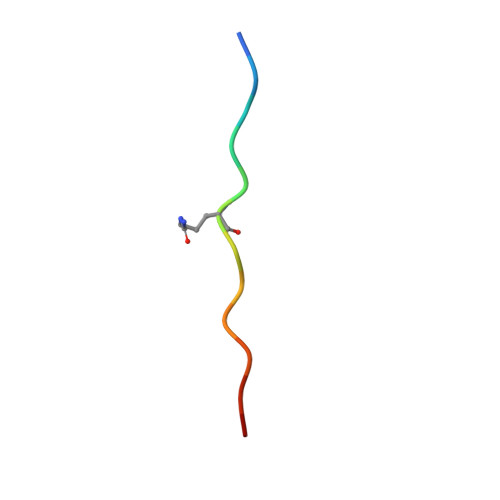
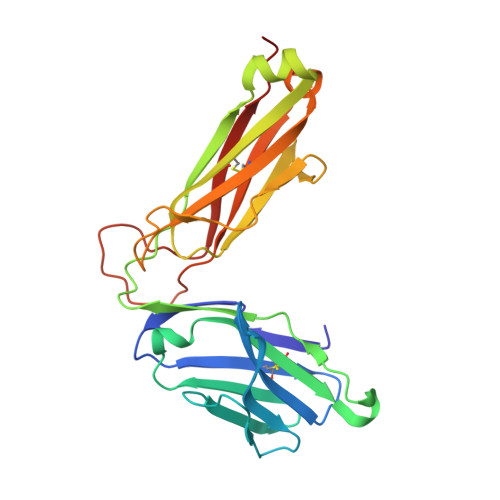
    CD4 + T cells recognising citrullinated self-epitopes presented by HLA-DRB1 bearing the shared susceptibility epitope (SE) are implicated in rheumatoid arthritis (RA). However, the underlying T cell receptor (TCR) determinants of epitope specificity towards distinct citrullinated peptide antigens, including vimentin-64cit 59-71 and α-enolase-15cit 10-22 remain unclear. Using HLA-DR4-tetramers, we examine the T cell repertoire in HLA-DR4 transgenic mice and observe biased TRAV6 TCR gene usage across these two citrullinated epitopes which matches with TCR bias previously observed towards the fibrinogen β-74cit 69-81 epitope. Moreover, shared TRAV26-1 gene usage is evident in four α-enolase-15cit 10-22 reactive T cells in three human samples. Crystal structures of mouse TRAV6 + and human TRAV26-1 + TCR-HLA-DR4 complexes presenting vimentin-64cit 59-71 and α-enolase-15cit 10-22 , respectively, show three-way interactions between the TCR, SE, citrulline, and the basis for the biased selection of TRAV genes. Position 2 of the citrullinated epitope is a key determinant underpinning TCR specificity. Accordingly, we provide a molecular basis of TCR specificity towards citrullinated epitopes.


  • Organizational Affiliation
    • Infection and Immunity Program and Department of Biochemistry and Molecular Biology, Biomedicine Discovery Institute, Monash University, Clayton, Australia.

Macromolecule Content 

  • Total Structure Weight: 194.49 kDa 
  • Atom Count: 13,047 
  • Modeled Residue Count: 1,613 
  • Deposited Residue Count: 1,672 
  • Unique protein chains: 5

Macromolecules

Find similar proteins by:|  3D Structure
Entity ID: 1
MoleculeChains  Sequence LengthOrganismDetailsImage
HLA class II histocompatibility antigen, DR alpha chain
A, F
181Homo sapiensMutation(s): 0 
Gene Names: HLA-DRAHLA-DRA1
UniProt & NIH Common Fund Data Resources
Find proteins for P01903 (Homo sapiens)
Explore P01903 
Go to UniProtKB:  P01903
PHAROS:  P01903
GTEx:  ENSG00000204287 
Entity Groups
Sequence Clusters30% Identity50% Identity70% Identity90% Identity95% Identity100% Identity
UniProt GroupP01903
Glycosylation
Glycosylation Sites: 2Go to GlyGen: P01903-1
Sequence Annotations
Expand
Reference Sequence
Find similar proteins by:|  3D Structure
Entity ID: 2
MoleculeChains  Sequence LengthOrganismDetailsImage
HLA class II histocompatibility antigen, DRB1 beta chain
B, G
190Homo sapiensMutation(s): 17 
Gene Names: HLA-DRB1
UniProt & NIH Common Fund Data Resources
Find proteins for P01911 (Homo sapiens)
Explore P01911 
Go to UniProtKB:  P01911
PHAROS:  P01911
GTEx:  ENSG00000196126 
Entity Groups
Sequence Clusters30% Identity50% Identity70% Identity90% Identity95% Identity100% Identity
UniProt GroupP01911
Glycosylation
Glycosylation Sites: 1Go to GlyGen: P01911-1
Sequence Annotations
Expand
Reference Sequence
Find similar proteins by:  Sequence   |   3D Structure  
Entity ID: 3
MoleculeChains  Sequence LengthOrganismDetailsImage
Vimentin
C, H
13Homo sapiensMutation(s): 0 
UniProt & NIH Common Fund Data Resources
Find proteins for P08670 (Homo sapiens)
Explore P08670 
Go to UniProtKB:  P08670
PHAROS:  P08670
GTEx:  ENSG00000026025 
Entity Groups
Sequence Clusters30% Identity50% Identity70% Identity90% Identity95% Identity100% Identity
UniProt GroupP08670
Sequence Annotations
Expand
Reference Sequence
Find similar proteins by:|  3D Structure
Entity ID: 4
MoleculeChains  Sequence LengthOrganismDetailsImage
A03 TCR alpha chain
D, I
209Mus musculusMutation(s): 0 
Find similar proteins by:|  3D Structure
Entity ID: 5
MoleculeChains  Sequence LengthOrganismDetailsImage
A03 TCR beta chain
E, J
243Mus musculusMutation(s): 0 

Oligosaccharides

Help  
Entity ID: 6
MoleculeChains Length2D Diagram GlycosylationD Interactions
2-acetamido-2-deoxy-beta-D-glucopyranose-(1-4)-2-acetamido-2-deoxy-beta-D-glucopyranose
K, L
2N-Glycosylation
Glycosylation Resources
GlyTouCan: G42666HT
GlyCosmos: G42666HT
GlyGen: G42666HT
Entity ID: 7
MoleculeChains Length2D Diagram GlycosylationD Interactions
2-acetamido-2-deoxy-beta-D-glucopyranose-(1-4)-2-acetamido-2-deoxy-beta-D-glucopyranose-(1-4)-2-acetamido-2-deoxy-beta-D-glucopyranose
M
3N-Glycosylation
Glycosylation Resources
GlyTouCan: G47362BJ
GlyCosmos: G47362BJ
GlyGen: G47362BJ

Small Molecules

Ligands 3 Unique
IDChains Name / Formula / InChI Key2D Diagram3D Interactions
NAG

Query on NAG


Download:Ideal Coordinates CCD File
BA [auth F],
S [auth B]
2-acetamido-2-deoxy-beta-D-glucopyranose
C8 H15 N O6
OVRNDRQMDRJTHS-FMDGEEDCSA-N
SO4

Query on SO4


Download:Ideal Coordinates CCD File
AA [auth E]
EA [auth F]
GA [auth G]
HA [auth G]
IA [auth G]
AA [auth E],
EA [auth F],
GA [auth G],
HA [auth G],
IA [auth G],
JA [auth G],
KA [auth G],
LA [auth I],
MA [auth I],
NA [auth J],
OA [auth J],
Q [auth A],
R [auth A],
V [auth B],
X [auth D],
Z [auth E]
SULFATE ION
O4 S
QAOWNCQODCNURD-UHFFFAOYSA-L
GOL

Query on GOL


Download:Ideal Coordinates CCD File
CA [auth F]
DA [auth F]
FA [auth G]
N [auth A]
O [auth A]
CA [auth F],
DA [auth F],
FA [auth G],
N [auth A],
O [auth A],
P [auth A],
T [auth B],
U [auth B],
W [auth D],
Y [auth E]
GLYCEROL
C3 H8 O3
PEDCQBHIVMGVHV-UHFFFAOYSA-N
Modified Residues  1 Unique
IDChains TypeFormula2D DiagramParent
CIR
Query on CIR
C, H
L-PEPTIDE LINKINGC6 H13 N3 O3ARG

Experimental Data & Validation

Experimental Data

  • Method: X-RAY DIFFRACTION
  • Resolution: 2.65 Å
  • R-Value Free:  0.244 (Depositor), 0.237 (DCC) 
  • R-Value Work:  0.209 (Depositor) 
  • R-Value Observed: 0.210 (Depositor) 
Space Group: I 1 2 1
Unit Cell:
Length ( Å )Angle ( ˚ )
a = 98.929α = 90
b = 59.97β = 90.16
c = 379.159γ = 90
Software Package:
Software NamePurpose
PHENIXrefinement
PHENIXrefinement
XDSdata reduction
Aimlessdata scaling
PHASERphasing

Structure Validation

View Full Validation Report


Ligand Structure Quality Assessment 


Entry History 

& Funding Information

Deposition Data


Funding OrganizationLocationGrant Number
National Health and Medical Research Council (NHMRC, Australia)Australia2008981
Janssen PharmaceuticalsUnited States--

Revision History  (Full details and data files)

  • Version 1.0: 2024-07-31
    Type: Initial release
  • Version 1.1: 2024-08-07
    Changes: Database references
  • Version 1.2: 2024-10-09
    Changes: Structure summary