8TM1

Antibody N3-1 bound to RBDs in the up and down conformations


Experimental Data Snapshot

  • Method: ELECTRON MICROSCOPY
  • Resolution: 2.79 Å
  • Aggregation State: PARTICLE 
  • Reconstruction Method: SINGLE PARTICLE 

wwPDB Validation   3D Report Full Report


This is version 1.0 of the entry. See complete history


Literature

SARS-COV-2 Omicron variants conformationally escape a rare quaternary antibody binding mode.

Goike, J.Hsieh, C.L.Horton, A.P.Gardner, E.C.Zhou, L.Bartzoka, F.Wang, N.Javanmardi, K.Herbert, A.Abbassi, S.Xie, X.Xia, H.Shi, P.Y.Renberg, R.Segall-Shapiro, T.H.Terrace, C.I.Wu, W.Shroff, R.Byrom, M.Ellington, A.D.Marcotte, E.M.Musser, J.M.Kuchipudi, S.V.Kapur, V.Georgiou, G.Weaver, S.C.Dye, J.M.Boutz, D.R.McLellan, J.S.Gollihar, J.D.

(2023) Commun Biol 6: 1250-1250

  • DOI: https://doi.org/10.1038/s42003-023-05649-6
  • Primary Citation of Related Structures:  
    8TM1, 8TMA

  • PubMed Abstract: 

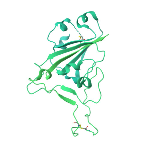
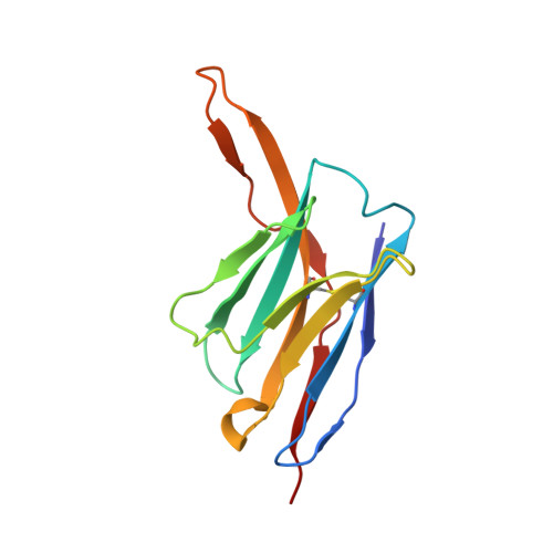
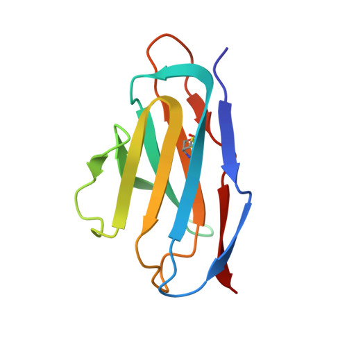
    The ongoing evolution of SARS-CoV-2 into more easily transmissible and infectious variants has provided unprecedented insight into mutations enabling immune escape. Understanding how these mutations affect the dynamics of antibody-antigen interactions is crucial to the development of broadly protective antibodies and vaccines. Here we report the characterization of a potent neutralizing antibody (N3-1) identified from a COVID-19 patient during the first disease wave. Cryogenic electron microscopy revealed a quaternary binding mode that enables direct interactions with all three receptor-binding domains of the spike protein trimer, resulting in extraordinary avidity and potent neutralization of all major variants of concern until the emergence of Omicron. Structure-based rational design of N3-1 mutants improved binding to all Omicron variants but only partially restored neutralization of the conformationally distinct Omicron BA.1. This study provides new insights into immune evasion through changes in spike protein dynamics and highlights considerations for future conformationally biased multivalent vaccine designs.


  • Organizational Affiliation

    Center for Systems and Synthetic Biology, Department of Molecular Biosciences, The University of Texas at Austin, Austin, TX, USA.


Macromolecules
Find similar proteins by:  (by identity cutoff)  |  3D Structure
Entity ID: 1
MoleculeChains Sequence LengthOrganismDetailsImage
Spike glycoprotein
A, B
1,288Severe acute respiratory syndrome coronavirus 2Mutation(s): 9 
Gene Names: S2
UniProt
Find proteins for P0DTC2 (Severe acute respiratory syndrome coronavirus 2)
Explore P0DTC2 
Go to UniProtKB:  P0DTC2
Entity Groups  
Sequence Clusters30% Identity50% Identity70% Identity90% Identity95% Identity100% Identity
UniProt GroupP0DTC2
Sequence Annotations
Expand
  • Reference Sequence
Find similar proteins by:  (by identity cutoff)  |  3D Structure
Entity ID: 2
MoleculeChains Sequence LengthOrganismDetailsImage
N3-1 Fab heavy chainC [auth H]128Homo sapiensMutation(s): 0 
Entity Groups  
Sequence Clusters30% Identity50% Identity70% Identity90% Identity95% Identity100% Identity
Sequence Annotations
Expand
  • Reference Sequence
Find similar proteins by:  (by identity cutoff)  |  3D Structure
Entity ID: 3
MoleculeChains Sequence LengthOrganismDetailsImage
N3-1 Fab light chainD [auth L]108Homo sapiensMutation(s): 0 
Entity Groups  
Sequence Clusters30% Identity50% Identity70% Identity90% Identity95% Identity100% Identity
Sequence Annotations
Expand
  • Reference Sequence
Experimental Data & Validation

Experimental Data

  • Method: ELECTRON MICROSCOPY
  • Resolution: 2.79 Å
  • Aggregation State: PARTICLE 
  • Reconstruction Method: SINGLE PARTICLE 

Structure Validation

View Full Validation Report


Entry History & Funding Information

Deposition Data


Funding OrganizationLocationGrant Number
National Institutes of Health/National Institute Of Allergy and Infectious Diseases (NIH/NIAID)United StatesR01 AI127521

Revision History  (Full details and data files)

  • Version 1.0: 2024-01-17
    Type: Initial release