New Insights into the Complement Receptor of the Ig Superfamily Obtained from Structural and Functional Studies on Two Mutants.
Duan, H., Abram, T.G., Cruz, A.R., Rooijakkers, S.H.M., Geisbrecht, B.V.(2023) Immunohorizons 7: 806-818
- PubMed: 38032267 Search on PubMedSearch on PubMed Central
- DOI: https://doi.org/10.4049/immunohorizons.2300064
- Primary Citation Related Structures:
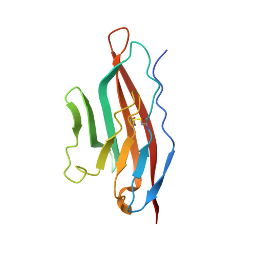
8TE5, 8TE6 - PubMed Abstract:
The extracellular region of the complement receptor of the Ig superfamily (CRIg) binds to certain C3 cleavage products (C3b, iC3b, C3c) and inhibits the alternative pathway (AP) of complement. In this study, we provide further insight into the CRIg protein and describe two CRIg mutants that lack multiple lysine residues as a means of facilitating chemical modifications of the protein. Structural analyses confirmed preservation of the native CRIg architecture in both mutants. In contrast to earlier reports suggesting that CRIg binds to C3b with an affinity of ∼1 μM, we found that wild-type CRIg binds to C3b and iC3b with affinities <100 nM, but to C3c with an affinity closer to 1 μM. We observed this same trend for both lysine substitution mutants, albeit with an apparent ∼2- to 3-fold loss of affinity when compared with wild-type CRIg. Using flow cytometry, we confirmed binding to C3 fragment-opsonized Staphylococcus aureus cells by each mutant, again with an ∼2- to 3-fold decrease when compared with wild-type. Whereas wild-type CRIg inhibits AP-driven lysis of rabbit erythrocytes with an IC50 of 1.6 μM, we observed an ∼3-fold reduction in inhibition for both mutants. Interestingly, we found that amine-reactive crosslinking of the CRIg mutant containing only a single lysine results in a significant improvement in inhibitory potency across all concentrations examined when compared with the unmodified mutant, but in a manner sensitive to the length of the crosslinker. Collectively, our findings provide new insights into the CRIg protein and suggest an approach for engineering increasingly potent CRIg-based inhibitors of the AP.
- Department of Biochemistry and Molecular Biophysics, Kansas State University; Manhattan, KS.
Organizational Affiliation: