8SZZ

CryoEM Structure of Computationally Designed Nanocage O32-ZL4


Experimental Data Snapshot

  • Method: ELECTRON MICROSCOPY
  • Resolution: 2.90 Å
  • Aggregation State: PARTICLE 
  • Reconstruction Method: SINGLE PARTICLE 

wwPDB Validation   3D Report Full Report


This is version 1.1 of the entry. See complete history


Literature

Accurate computational design of three-dimensional protein crystals.

Li, Z.Wang, S.Nattermann, U.Bera, A.K.Borst, A.J.Yaman, M.Y.Bick, M.J.Yang, E.C.Sheffler, W.Lee, B.Seifert, S.Hura, G.L.Nguyen, H.Kang, A.Dalal, R.Lubner, J.M.Hsia, Y.Haddox, H.Courbet, A.Dowling, Q.Miranda, M.Favor, A.Etemadi, A.Edman, N.I.Yang, W.Weidle, C.Sankaran, B.Negahdari, B.Ross, M.B.Ginger, D.S.Baker, D.

(2023) Nat Mater 22: 1556-1563

  • DOI: https://doi.org/10.1038/s41563-023-01683-1
  • Primary Citation of Related Structures:  
    8CUS, 8CUT, 8CUU, 8CUV, 8CUW, 8CUX, 8CWS, 8CWY, 8CWZ, 8FAR, 8SZZ

  • PubMed Abstract: 

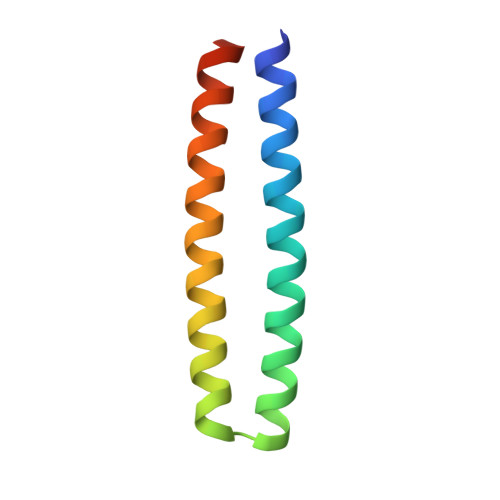
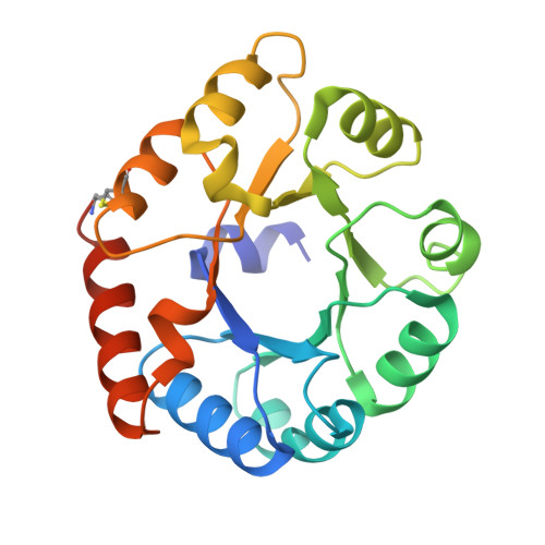
    Protein crystallization plays a central role in structural biology. Despite this, the process of crystallization remains poorly understood and highly empirical, with crystal contacts, lattice packing arrangements and space group preferences being largely unpredictable. Programming protein crystallization through precisely engineered side-chain-side-chain interactions across protein-protein interfaces is an outstanding challenge. Here we develop a general computational approach for designing three-dimensional protein crystals with prespecified lattice architectures at atomic accuracy that hierarchically constrains the overall number of degrees of freedom of the system. We design three pairs of oligomers that can be individually purified, and upon mixing, spontaneously self-assemble into >100 µm three-dimensional crystals. The structures of these crystals are nearly identical to the computational design models, closely corresponding in both overall architecture and the specific protein-protein interactions. The dimensions of the crystal unit cell can be systematically redesigned while retaining the space group symmetry and overall architecture, and the crystals are extremely porous and highly stable. Our approach enables the computational design of protein crystals with high accuracy, and the designed protein crystals, which have both structural and assembly information encoded in their primary sequences, provide a powerful platform for biological materials engineering.


  • Organizational Affiliation

    Department of Biochemistry, University of Washington, Seattle, WA, USA.


Macromolecules
Find similar proteins by:  (by identity cutoff)  |  3D Structure
Entity ID: 1
MoleculeChains Sequence LengthOrganismDetailsImage
O32-ZL4 Component A76synthetic constructMutation(s): 0 
Entity Groups  
Sequence Clusters30% Identity50% Identity70% Identity90% Identity95% Identity100% Identity
Sequence Annotations
Expand
  • Reference Sequence
Find similar proteins by:  (by identity cutoff)  |  3D Structure
Entity ID: 2
MoleculeChains Sequence LengthOrganismDetailsImage
O32-ZL4 Component B213Thermotoga maritimaMutation(s): 0 
Gene Names: TM_0066
UniProt
Find proteins for Q9WXS1 (Thermotoga maritima (strain ATCC 43589 / DSM 3109 / JCM 10099 / NBRC 100826 / MSB8))
Explore Q9WXS1 
Go to UniProtKB:  Q9WXS1
Entity Groups  
Sequence Clusters30% Identity50% Identity70% Identity90% Identity95% Identity100% Identity
UniProt GroupQ9WXS1
Sequence Annotations
Expand
  • Reference Sequence
Small Molecules
Ligands 1 Unique
IDChains Name / Formula / InChI Key2D Diagram3D Interactions
NA
Query on NA

Download Ideal Coordinates CCD File 
AB [auth M]
BB [auth N]
CB [auth O]
DB [auth U]
EB [auth Y]
AB [auth M],
BB [auth N],
CB [auth O],
DB [auth U],
EB [auth Y],
FB [auth Z],
GB [auth a],
HB [auth b],
IB [auth c],
JB [auth d],
KB [auth e],
LB [auth f],
MB [auth g],
NB [auth h],
OB [auth i],
PB [auth j],
QB [auth k],
RB [auth l],
SB [auth m],
TB [auth n],
WA [auth I],
XA [auth J],
YA [auth K],
ZA [auth L]
SODIUM ION
Na
FKNQFGJONOIPTF-UHFFFAOYSA-N
Experimental Data & Validation

Experimental Data

  • Method: ELECTRON MICROSCOPY
  • Resolution: 2.90 Å
  • Aggregation State: PARTICLE 
  • Reconstruction Method: SINGLE PARTICLE 
EM Software:
TaskSoftware PackageVersion
MODEL REFINEMENTCoot
MODEL REFINEMENTPHENIX
MODEL REFINEMENTRosetta

Structure Validation

View Full Validation Report


Entry History & Funding Information

Deposition Data


Funding OrganizationLocationGrant Number
Howard Hughes Medical Institute (HHMI)United States--

Revision History  (Full details and data files)

  • Version 1.0: 2023-11-01
    Type: Initial release
  • Version 1.1: 2023-12-27
    Changes: Database references