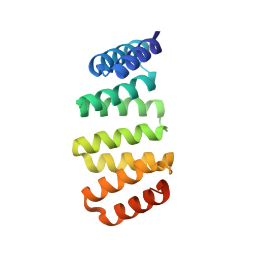
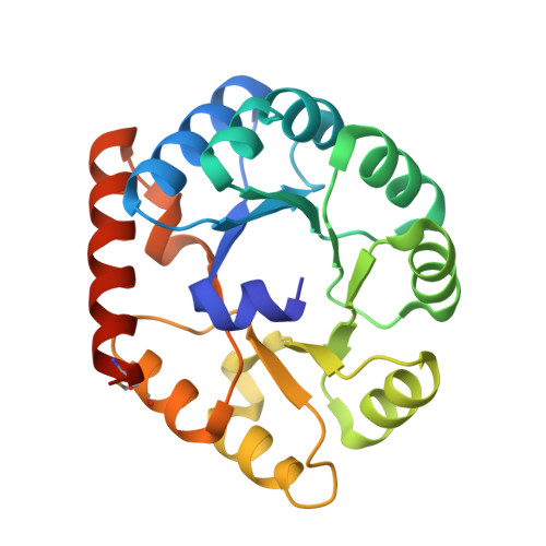
Accurate computational design of three-dimensional protein crystals.
Li, Z., Wang, S., Nattermann, U., Bera, A.K., Borst, A.J., Yaman, M.Y., Bick, M.J., Yang, E.C., Sheffler, W., Lee, B., Seifert, S., Hura, G.L., Nguyen, H., Kang, A., Dalal, R., Lubner, J.M., Hsia, Y., Haddox, H., Courbet, A., Dowling, Q., Miranda, M., Favor, A., Etemadi, A., Edman, N.I., Yang, W., Weidle, C., Sankaran, B., Negahdari, B., Ross, M.B., Ginger, D.S., Baker, D.(2023) Nat Mater 22: 1556-1563
- PubMed: 37845322
- DOI: https://doi.org/10.1038/s41563-023-01683-1
- Primary Citation of Related Structures:
8CUS, 8CUT, 8CUU, 8CUV, 8CUW, 8CUX, 8CWS, 8CWY, 8CWZ, 8FAR, 8SZZ - PubMed Abstract:
Protein crystallization plays a central role in structural biology. Despite this, the process of crystallization remains poorly understood and highly empirical, with crystal contacts, lattice packing arrangements and space group preferences being largely unpredictable. Programming protein crystallization through precisely engineered side-chain-side-chain interactions across protein-protein interfaces is an outstanding challenge. Here we develop a general computational approach for designing three-dimensional protein crystals with prespecified lattice architectures at atomic accuracy that hierarchically constrains the overall number of degrees of freedom of the system. We design three pairs of oligomers that can be individually purified, and upon mixing, spontaneously self-assemble into >100 µm three-dimensional crystals. The structures of these crystals are nearly identical to the computational design models, closely corresponding in both overall architecture and the specific protein-protein interactions. The dimensions of the crystal unit cell can be systematically redesigned while retaining the space group symmetry and overall architecture, and the crystals are extremely porous and highly stable. Our approach enables the computational design of protein crystals with high accuracy, and the designed protein crystals, which have both structural and assembly information encoded in their primary sequences, provide a powerful platform for biological materials engineering.
- Department of Biochemistry, University of Washington, Seattle, WA, USA.
Organizational Affiliation:


