8STD

S127A variant of LarB, a carboxylase/hydrolase involved in synthesis of the cofactor for lactate racemase, in complex with authentic substrate NaAD and soaked with CS2


Experimental Data Snapshot

  • Method: X-RAY DIFFRACTION
  • Resolution: 2.65 Å
  • R-Value Free: 0.273 
  • R-Value Work: 0.242 
  • R-Value Observed: 0.243 

wwPDB Validation   3D Report Full Report


Ligand Structure Quality Assessment 


This is version 1.0 of the entry. See complete history


Literature

Structure of the LarB-Substrate Complex and Identification of a Reaction Intermediate during Nickel-Pincer Nucleotide Cofactor Biosynthesis.

Chatterjee, S.Nevarez, J.L.Rankin, J.A.Hu, J.Hausinger, R.P.

(2023) Biochemistry 62: 3096-3104

  • DOI: https://doi.org/10.1021/acs.biochem.3c00242
  • Primary Citation of Related Structures:  
    8SOQ, 8STD

  • PubMed Abstract: 

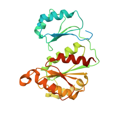
    LarB catalyzes the first step of biosynthesis for the nickel-pincer nucleotide cofactor by converting nicotinic acid adenine dinucleotide (NaAD) to AMP and pyridinium-3,5-biscarboxylic acid mononucleotide (P2CMN). Prior studies had shown that LarB uses CO 2 for substrate carboxylation and reported the structure of a Lactiplantibacillus plantarum LarB·NAD + complex, revealing a covalent linkage between Cys221 and C4 of the pyridine ring. This interaction was proposed to promote C5 carboxylation, with C5-carboxylated-NaAD suggested to activate magnesium-bound water, leading to phosphoanhydride hydrolysis. Here, we extended the analysis of wild-type LarB by using ultraviolet-visible spectroscopy to obtain additional evidence for cysteinyl side chain attachment to the ring of NAD + , thus demonstrating that this linkage is not a crystallization artifact. Using the S127A variant of L. plantarum LarB, a form of the enzyme with a reduced rate of NaAD hydrolysis, we examined its interaction with the authentic substrate. The intermediate arising from C5 carboxylation of NaAD, dinicotinic acid adenine dinucleotide (DaAD), was identified by using mass spectrometry. S127A LarB exhibited spectroscopic evidence of a Cys221-NAD + adduct, but a covalent enzyme-NaAD linkage was not detectable. We determined the S127A LarB·NaAD structure, providing new insights into the enzyme mechanism, and tentatively identified the position and mode of CO 2 binding. The crystal structure revealed the location of the side chain for Glu180, which was previously disordered, but showed that it is not well positioned to abstract the C5 proton in the adduct species to restore aromaticity as Cys221 is expelled. Based on these combined results, we propose a revised catalytic mechanism of LarB..


  • Organizational Affiliation

    Department of Microbiology and Molecular Genetics, Michigan State University, East Lansing, Michigan 48824, United States.


Macromolecules
Find similar proteins by:  (by identity cutoff)  |  3D Structure
Entity ID: 1
MoleculeChains Sequence LengthOrganismDetailsImage
Pyridinium-3,5-biscarboxylic acid mononucleotide synthase
A, B, C, D, E
A, B, C, D, E, F
256Lactiplantibacillus plantarum WCFS1Mutation(s): 0 
Gene Names: larBlp_0105
EC: 2.5.1.143
UniProt
Find proteins for F9UST0 (Lactiplantibacillus plantarum (strain ATCC BAA-793 / NCIMB 8826 / WCFS1))
Explore F9UST0 
Go to UniProtKB:  F9UST0
Entity Groups  
Sequence Clusters30% Identity50% Identity70% Identity90% Identity95% Identity100% Identity
UniProt GroupF9UST0
Sequence Annotations
Expand
  • Reference Sequence
Small Molecules
Ligands 2 Unique
IDChains Name / Formula / InChI Key2D Diagram3D Interactions
DND (Subject of Investigation/LOI)
Query on DND

Download Ideal Coordinates CCD File 
I [auth A]
L [auth B]
O [auth C]
Q [auth D]
S [auth E]
I [auth A],
L [auth B],
O [auth C],
Q [auth D],
S [auth E],
U [auth F]
NICOTINIC ACID ADENINE DINUCLEOTIDE
C21 H27 N6 O15 P2
SENPVEZBRZQVST-HISDBWNOSA-O
MG
Query on MG

Download Ideal Coordinates CCD File 
G [auth A]
H [auth A]
J [auth B]
K [auth B]
M [auth C]
G [auth A],
H [auth A],
J [auth B],
K [auth B],
M [auth C],
N [auth C],
P [auth D],
R [auth E],
T [auth F]
MAGNESIUM ION
Mg
JLVVSXFLKOJNIY-UHFFFAOYSA-N
Experimental Data & Validation

Experimental Data

  • Method: X-RAY DIFFRACTION
  • Resolution: 2.65 Å
  • R-Value Free: 0.273 
  • R-Value Work: 0.242 
  • R-Value Observed: 0.243 
  • Space Group: P 4 21 2
Unit Cell:
Length ( Å )Angle ( ˚ )
a = 118.839α = 90
b = 118.839β = 90
c = 211.289γ = 90
Software Package:
Software NamePurpose
PHENIXrefinement
iMOSFLMdata reduction
Aimlessdata scaling
PHASERphasing

Structure Validation

View Full Validation Report


Ligand Structure Quality Assessment 


Entry History & Funding Information

Deposition Data


Funding OrganizationLocationGrant Number
National Science Foundation (NSF, United States)United StatesCHE-1807073
National Institutes of Health/National Institute of General Medical Sciences (NIH/NIGMS)United StatesGM128959

Revision History  (Full details and data files)

  • Version 1.0: 2023-12-27
    Type: Initial release