8SP0

Symmetric dimer of MapSPARTA bound with gRNA/tDNA hybrid


Experimental Data Snapshot

  • Method: ELECTRON MICROSCOPY
  • Resolution: 3.33 Å
  • Aggregation State: PARTICLE 
  • Reconstruction Method: SINGLE PARTICLE 

wwPDB Validation   3D Report Full Report


This is version 1.1 of the entry. See complete history


Literature

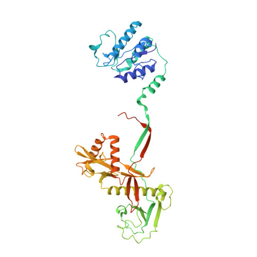
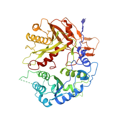
Oligomerization-mediated activation of a short prokaryotic Argonaute.

Shen, Z.Yang, X.Y.Xia, S.Huang, W.Taylor, D.J.Nakanishi, K.Fu, T.M.

(2023) Nature 621: 154-161

  • DOI: https://doi.org/10.1038/s41586-023-06456-z
  • Primary Citation of Related Structures:  
    8FEX, 8FFI, 8SP0, 8SP3, 8SPO, 8SQU

  • PubMed Abstract: 

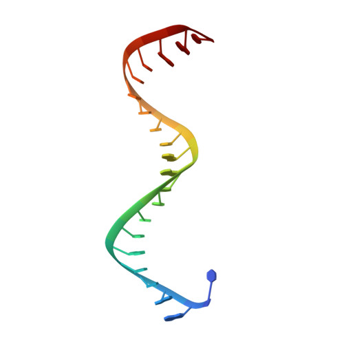
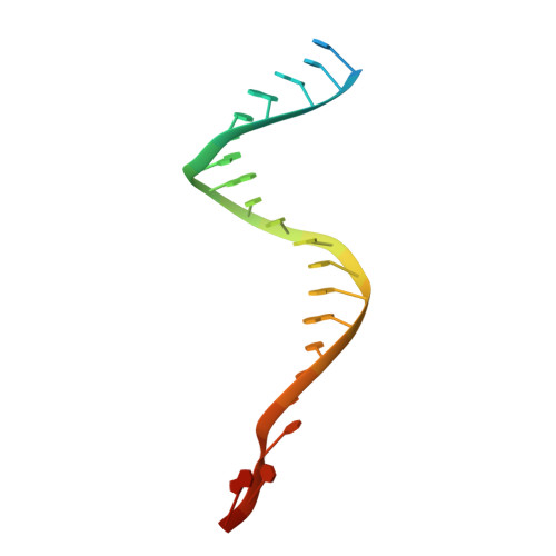
    Although eukaryotic and long prokaryotic Argonaute proteins (pAgos) cleave nucleic acids, some short pAgos lack nuclease activity and hydrolyse NAD(P) + to induce bacterial cell death 1 . Here we present a hierarchical activation pathway for SPARTA, a short pAgo consisting of an Argonaute (Ago) protein and TIR-APAZ, an associated protein 2 . SPARTA progresses through distinct oligomeric forms, including a monomeric apo state, a monomeric RNA-DNA-bound state, two dimeric RNA-DNA-bound states and a tetrameric RNA-DNA-bound active state. These snapshots together identify oligomerization as a mechanistic principle of SPARTA activation. The RNA-DNA-binding channel of apo inactive SPARTA is occupied by an auto-inhibitory motif in TIR-APAZ. After the binding of RNA-DNA, SPARTA transitions from a monomer to a symmetric dimer and then an asymmetric dimer, in which two TIR domains interact through charge and shape complementarity. Next, two dimers assemble into a tetramer with a central TIR cluster responsible for hydrolysing NAD(P) + . In addition, we observe unique features of interactions between SPARTA and RNA-DNA, including competition between the DNA 3' end and the auto-inhibitory motif, interactions between the RNA G2 nucleotide and Ago, and splaying of the RNA-DNA duplex by two loops exclusive to short pAgos. Together, our findings provide a mechanistic basis for the activation of short pAgos, a large section of the Ago superfamily.


  • Organizational Affiliation

    Department of Biological Chemistry and Pharmacology, The Ohio State University, Columbus, OH, USA.


Macromolecules

Find similar proteins by:  (by identity cutoff)  |  3D Structure
Entity ID: 1
MoleculeChains Sequence LengthOrganismDetailsImage
TIR-APAZ
A, E
451Maribacter polysiphoniaeMutation(s): 0 
Gene Names: LX92_01810
UniProt
Find proteins for A0A316E683 (Maribacter polysiphoniae)
Explore A0A316E683 
Go to UniProtKB:  A0A316E683
Entity Groups  
Sequence Clusters30% Identity50% Identity70% Identity90% Identity95% Identity100% Identity
UniProt GroupA0A316E683
Sequence Annotations
Expand
  • Reference Sequence
Find similar proteins by:  (by identity cutoff)  |  3D Structure
Entity ID: 2
MoleculeChains Sequence LengthOrganismDetailsImage
short pAgo
B, F
507Maribacter polysiphoniaeMutation(s): 0 
Gene Names: LX92_01809
UniProt
Find proteins for A0A316E3U6 (Maribacter polysiphoniae)
Explore A0A316E3U6 
Go to UniProtKB:  A0A316E3U6
Entity Groups  
Sequence Clusters30% Identity50% Identity70% Identity90% Identity95% Identity100% Identity
UniProt GroupA0A316E3U6
Sequence Annotations
Expand
  • Reference Sequence

Find similar nucleic acids by:  Sequence   |   3D Structure  

Entity ID: 3
MoleculeChains LengthOrganismImage
guide RNA
C, G
21synthetic construct
Sequence Annotations
Expand
  • Reference Sequence

Find similar nucleic acids by:  Sequence   |   3D Structure  

Entity ID: 4
MoleculeChains LengthOrganismImage
target DNA
D, H
25synthetic construct
Sequence Annotations
Expand
  • Reference Sequence
Experimental Data & Validation

Experimental Data

  • Method: ELECTRON MICROSCOPY
  • Resolution: 3.33 Å
  • Aggregation State: PARTICLE 
  • Reconstruction Method: SINGLE PARTICLE 

Structure Validation

View Full Validation Report


Entry History & Funding Information

Deposition Data


Funding OrganizationLocationGrant Number
Not funded--

Revision History  (Full details and data files)

  • Version 1.0: 2023-08-23
    Type: Initial release
  • Version 1.1: 2023-09-20
    Changes: Database references