8SK7

Cryo-EM structure of designed Influenza HA binder, HA_20, bound to Influenza HA (Strain: Iowa43)


Experimental Data Snapshot

  • Method: ELECTRON MICROSCOPY
  • Resolution: 2.93 Å
  • Aggregation State: PARTICLE 
  • Reconstruction Method: SINGLE PARTICLE 

wwPDB Validation   3D Report Full Report


This is version 1.3 of the entry. See complete history


Literature

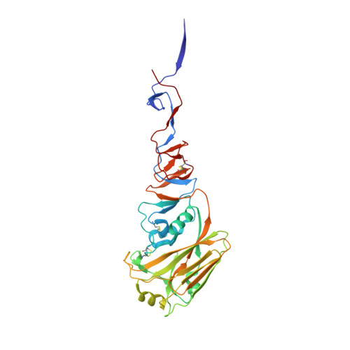
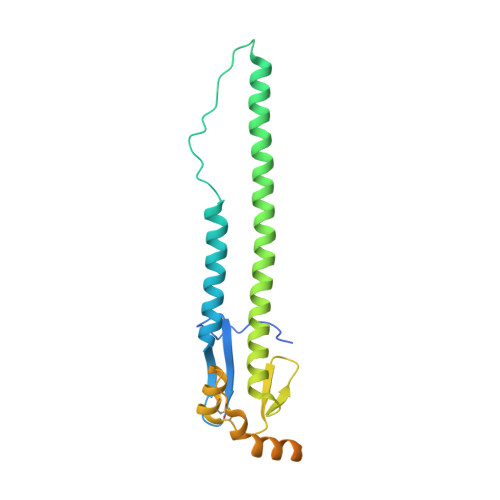
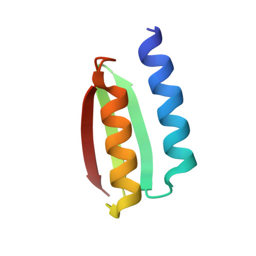
De novo design of protein structure and function with RFdiffusion.

Watson, J.L.Juergens, D.Bennett, N.R.Trippe, B.L.Yim, J.Eisenach, H.E.Ahern, W.Borst, A.J.Ragotte, R.J.Milles, L.F.Wicky, B.I.M.Hanikel, N.Pellock, S.J.Courbet, A.Sheffler, W.Wang, J.Venkatesh, P.Sappington, I.Torres, S.V.Lauko, A.De Bortoli, V.Mathieu, E.Ovchinnikov, S.Barzilay, R.Jaakkola, T.S.DiMaio, F.Baek, M.Baker, D.

(2023) Nature 620: 1089-1100

  • DOI: https://doi.org/10.1038/s41586-023-06415-8
  • Primary Citation of Related Structures:  
    8SK7

  • PubMed Abstract: 

    There has been considerable recent progress in designing new proteins using deep-learning methods 1-9 . Despite this progress, a general deep-learning framework for protein design that enables solution of a wide range of design challenges, including de novo binder design and design of higher-order symmetric architectures, has yet to be described. Diffusion models 10,11 have had considerable success in image and language generative modelling but limited success when applied to protein modelling, probably due to the complexity of protein backbone geometry and sequence-structure relationships. Here we show that by fine-tuning the RoseTTAFold structure prediction network on protein structure denoising tasks, we obtain a generative model of protein backbones that achieves outstanding performance on unconditional and topology-constrained protein monomer design, protein binder design, symmetric oligomer design, enzyme active site scaffolding and symmetric motif scaffolding for therapeutic and metal-binding protein design. We demonstrate the power and generality of the method, called RoseTTAFold diffusion (RFdiffusion), by experimentally characterizing the structures and functions of hundreds of designed symmetric assemblies, metal-binding proteins and protein binders. The accuracy of RFdiffusion is confirmed by the cryogenic electron microscopy structure of a designed binder in complex with influenza haemagglutinin that is nearly identical to the design model. In a manner analogous to networks that produce images from user-specified inputs, RFdiffusion enables the design of diverse functional proteins from simple molecular specifications.


  • Organizational Affiliation

    Department of Biochemistry, University of Washington, Seattle, WA, USA.


Macromolecules
Find similar proteins by:  (by identity cutoff)  |  3D Structure
Entity ID: 1
MoleculeChains Sequence LengthOrganismDetailsImage
Hemagglutinin HA1 chainA,
D [auth B],
G [auth C]
321Influenza A virusMutation(s): 0 
Gene Names: HA
UniProt
Find proteins for A4GCK8 (Influenza A virus (strain A/USA:Iowa/1943 H1N1))
Explore A4GCK8 
Go to UniProtKB:  A4GCK8
Entity Groups  
Sequence Clusters30% Identity50% Identity70% Identity90% Identity95% Identity100% Identity
UniProt GroupA4GCK8
Sequence Annotations
Expand
  • Reference Sequence
Find similar proteins by:  (by identity cutoff)  |  3D Structure
Entity ID: 2
MoleculeChains Sequence LengthOrganismDetailsImage
HemagglutininB [auth G],
E [auth H],
H [auth I]
232Influenza A virusMutation(s): 0 
Gene Names: HA
UniProt
Find proteins for A4GCK8 (Influenza A virus (strain A/USA:Iowa/1943 H1N1))
Explore A4GCK8 
Go to UniProtKB:  A4GCK8
Entity Groups  
Sequence Clusters30% Identity50% Identity70% Identity90% Identity95% Identity100% Identity
UniProt GroupA4GCK8
Sequence Annotations
Expand
  • Reference Sequence
Find similar proteins by:  (by identity cutoff)  |  3D Structure
Entity ID: 3
MoleculeChains Sequence LengthOrganismDetailsImage
HA_20 minibinder (RFdiffusion-designed)C [auth X],
F [auth Y],
I [auth Z]
65unidentifiedMutation(s): 0 
Entity Groups  
Sequence Clusters30% Identity50% Identity70% Identity90% Identity95% Identity100% Identity
Sequence Annotations
Expand
  • Reference Sequence
Oligosaccharides

Help

Entity ID: 4
MoleculeChains Length2D Diagram Glycosylation3D Interactions
beta-D-mannopyranose-(1-4)-2-acetamido-2-deoxy-beta-D-glucopyranose-(1-4)-2-acetamido-2-deoxy-beta-D-glucopyranoseJ [auth D],
L [auth F],
N [auth K]
3N-Glycosylation
Glycosylation Resources
GlyTouCan:  G15407YE
GlyCosmos:  G15407YE
GlyGen:  G15407YE
Entity ID: 5
MoleculeChains Length2D Diagram Glycosylation3D Interactions
2-acetamido-2-deoxy-beta-D-glucopyranose-(1-4)-2-acetamido-2-deoxy-beta-D-glucopyranoseK [auth E],
M [auth J],
O [auth L]
2N-Glycosylation
Glycosylation Resources
GlyTouCan:  G42666HT
GlyCosmos:  G42666HT
GlyGen:  G42666HT
Small Molecules
Ligands 1 Unique
IDChains Name / Formula / InChI Key2D Diagram3D Interactions
NAG (Subject of Investigation/LOI)
Query on NAG

Download Ideal Coordinates CCD File 
AA [auth C]
P [auth A]
Q [auth A]
R [auth A]
S [auth A]
AA [auth C],
P [auth A],
Q [auth A],
R [auth A],
S [auth A],
T [auth B],
U [auth B],
V [auth B],
W [auth B],
X [auth C],
Y [auth C],
Z [auth C]
2-acetamido-2-deoxy-beta-D-glucopyranose
C8 H15 N O6
OVRNDRQMDRJTHS-FMDGEEDCSA-N
Experimental Data & Validation

Experimental Data

  • Method: ELECTRON MICROSCOPY
  • Resolution: 2.93 Å
  • Aggregation State: PARTICLE 
  • Reconstruction Method: SINGLE PARTICLE 

Structure Validation

View Full Validation Report


Entry History & Funding Information

Deposition Data


Funding OrganizationLocationGrant Number
Howard Hughes Medical Institute (HHMI)United States--

Revision History  (Full details and data files)

  • Version 1.0: 2023-06-14
    Type: Initial release
  • Version 1.1: 2023-07-12
    Changes: Database references
  • Version 1.2: 2023-07-26
    Changes: Database references
  • Version 1.3: 2023-09-13
    Changes: Data collection, Database references