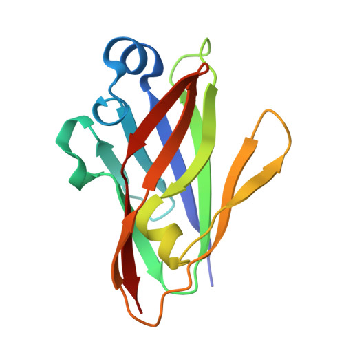
MakC and MakD are two proteins associated with a tripartite toxin of Vibrio cholerae .
Bodra, N., Toh, E., Nadeem, A., Wai, S.N., Persson, K.(2024) Front Microbiol 15: 1457850-1457850
- PubMed: 39421563 Search on PubMedSearch on PubMed Central
- DOI: https://doi.org/10.3389/fmicb.2024.1457850
- Primary Citation Related Structures:
8RQY - PubMed Abstract:
Pathogenic serotypes of Vibrio cholerae , transmitted through contaminated water and food, are responsible for outbreaks of cholera, an acute diarrheal disease. While the cholera toxin is the primary virulence factor, V. cholerae also expresses other virulence factors, such as the tripartite toxin MakABE that is secreted via the bacterial flagellum. These three proteins are co-expressed with two accessory proteins, MakC and MakD, whose functions remain unknown. Here, we present the crystal structures of MakC and MakD, revealing that they are similar in both sequence and structure but lack other close structural relatives. Our study further investigates the roles of MakC and MakD, focusing on their impact on the expression and secretion of the components of the MakABE tripartite toxin. Through deletion mutant analysis, we found that individual deletions of makC or makD do not significantly affect MakA expression or secretion. However, the deletion of both makC and makD impairs the expression of MakB, which is directly downstream, and decreases the expression of MakE, which is separated from makCD by two genes. Conversely, MakA, encoded by the makA gene located between makB and makE, is expressed normally but its secretion is impaired. Additionally, our findings indicate that MakC, in contrast to MakD, exhibits strong interactions with other proteins. Furthermore, both MakC and MakD were observed to be localized within the cytosol of the bacterial cell. This study provides new insights into the regulatory mechanisms affecting the Mak protein family in V. cholerae and highlights the complex interplay between gene proximity and protein expression.
- Department of Chemistry, Umeå University, Umeå, Sweden.
Organizational Affiliation: