8PKC | pdb_00008pkc

Structure of Api m1 in complex with the AM1-4 nanobody


Experimental Data Snapshot

  • Method: X-RAY DIFFRACTION
  • Resolution: 4.10 Å
  • R-Value Free: 
    0.296 (Depositor), 0.295 (DCC) 
  • R-Value Work: 
    0.258 (Depositor), 0.258 (DCC) 
  • R-Value Observed: 
    0.262 (Depositor) 

Starting Model: experimental
View more details

wwPDB Validation 3D Report Full Report

Validation slider image for 8PKC

Ligand Structure Quality Assessment 


This is version 1.2 of the entry. See complete history

Literature

Nanobody-based IgG simultaneously inhibit the allergenic and enzymatic activity of the dominant honeybee venom allergen.

Aagaard, J.B.Gandini, R.Ballegaard, A.R.Jensen, B.K.Dorn, B.Larsen, A.V.B.Lenstrup, A.Sonderholm, M.Miehe, M.Pfutzner, W.Jakob, T.Andersen, G.R.Mobs, C.Spillner, E.

(2026) Nat Commun 17: 1814-1814

  • DOI: https://doi.org/10.1038/s41467-026-69572-0
  • Primary Citation Related Structures: 
    8PIH, 8PKC

  • PubMed Abstract: 

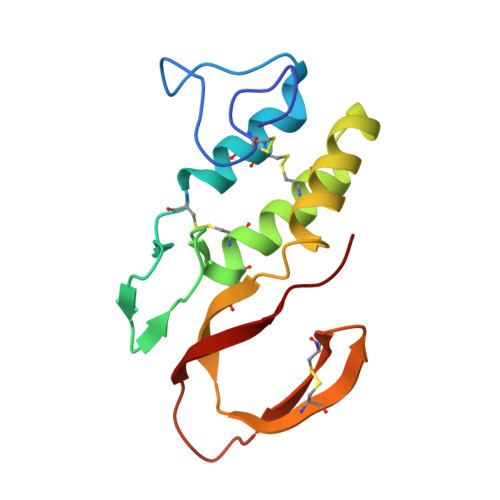
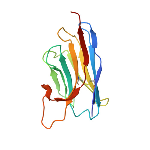
    Insect venoms can cause severe allergic reactions, including anaphylaxis, in sensitized individuals. In this study, we aim at preventing anaphylaxis mediated by the most abundant and dominant honeybee venom allergen phospholipase A2 (Api m 1) by blocking its interaction with allergic patient IgE. Therefore, we characterize selected Api m 1-specific nanobodies and identify two high-affinity binders with non-overlapping epitopes. Crystal structures of Api m 1/nanobody complexes reveal diametrically opposed epitopes, one of which involves the active site of Api m 1. Based on this background, we develop mono- and bispecific nanobody-human IgG 1 Fc, which exhibits pronounced blocking of IgE binding and effector cell activation in blood samples from honeybee venom allergic patients and reduces systemic reactions in a mouse model of allergen-induced anaphylaxis. This work provides a rationale for using nanobody-based inhibitors to prevent Api m 1-mediated anaphylaxis in honeybee venom allergy.


  • Organizational Affiliation
    • Department of Biological and Chemical Engineering, Aarhus University, Aarhus, Denmark.

Macromolecule Content 

  • Total Structure Weight: 58.58 kDa 
  • Atom Count: 4,065 
  • Modeled Residue Count: 522 
  • Deposited Residue Count: 526 
  • Unique protein chains: 2

Macromolecules

Find similar proteins by:|  3D Structure
Entity ID: 1
MoleculeChains  Sequence LengthOrganismDetailsImage
Phospholipase A2
A, B
134Apis melliferaMutation(s): 0 
EC: 3.1.1.4
UniProt
Find proteins for P00630 (Apis mellifera)
Explore P00630 
Go to UniProtKB:  P00630
Entity Groups
Sequence Clusters30% Identity50% Identity70% Identity90% Identity95% Identity100% Identity
UniProt GroupP00630
Glycosylation
Glycosylation Sites: 1
Sequence Annotations
Expand
Reference Sequence
Find similar proteins by:|  3D Structure
Entity ID: 2
MoleculeChains  Sequence LengthOrganismDetailsImage
AM1-4 nanobody
C, D
129Lama glamaMutation(s): 0 

Experimental Data & Validation

Experimental Data

  • Method: X-RAY DIFFRACTION
  • Resolution: 4.10 Å
  • R-Value Free:  0.296 (Depositor), 0.295 (DCC) 
  • R-Value Work:  0.258 (Depositor), 0.258 (DCC) 
  • R-Value Observed: 0.262 (Depositor) 
Space Group: P 41 21 2
Unit Cell:
Length ( Å )Angle ( ˚ )
a = 93.6α = 90
b = 93.6β = 90
c = 189.49γ = 90
Software Package:
Software NamePurpose
PHENIXrefinement
PHENIXrefinement
XDSdata reduction
XSCALEdata scaling
PHASERphasing

Structure Validation

View Full Validation Report


Ligand Structure Quality Assessment 


Entry History 

& Funding Information

Deposition Data


Funding OrganizationLocationGrant Number
Danish Council for Independent ResearchDenmark0135-00061B

Revision History  (Full details and data files)

  • Version 1.0: 2024-05-08
    Type: Initial release
  • Version 1.1: 2024-11-13
    Changes: Structure summary
  • Version 1.2: 2026-07-01
    Changes: Database references