8OGI

Structure of native human eosinophil peroxidase


Experimental Data Snapshot

  • Method: X-RAY DIFFRACTION
  • Resolution: 1.55 Å
  • R-Value Free: 0.185 
  • R-Value Work: 0.174 
  • R-Value Observed: 0.175 

wwPDB Validation   3D Report Full Report


Ligand Structure Quality Assessment 


This is version 1.1 of the entry. See complete history


Literature

Posttranslational modification and heme cavity architecture of human eosinophil peroxidase-insights from first crystal structure and biochemical characterization.

Pfanzagl, V.Gruber-Grunwald, C.Leitgeb, U.Furtmuller, P.G.Obinger, C.

(2023) J Biol Chem 299: 105402-105402

  • DOI: https://doi.org/10.1016/j.jbc.2023.105402
  • Primary Citation of Related Structures:  
    8OGI

  • PubMed Abstract: 

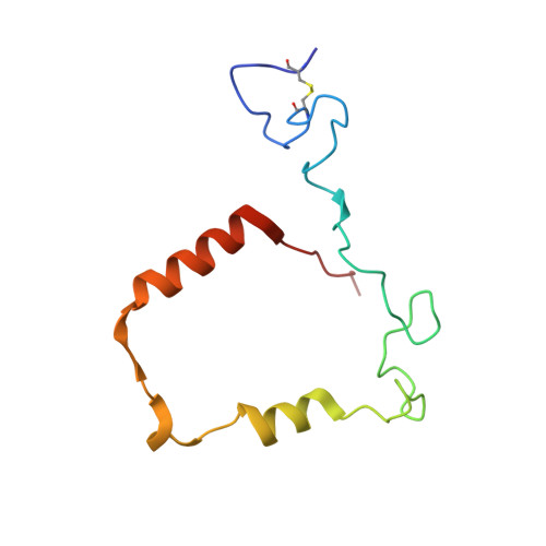
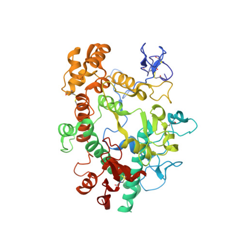
    Eosinophil peroxidase (EPO) is the most abundant granule protein exocytosed by eosinophils, specialized human phagocytes. Released EPO catalyzes the formation of reactive oxidants from bromide, thiocyanate, and nitrite that kill tissue-invading parasites. However, EPO also plays a deleterious role in inflammatory diseases, making it a potential pharmacological target. A major hurdle is the high similarity to the homologous myeloperoxidase (MPO), which requires a detailed understanding of the small structural differences that can be used to increase the specificity of the inhibitors. Here, we present the first crystal structure of mature leukocyte EPO at 1.6 Å resolution together with analyses of its posttranslational modifications and biochemical properties. EPO has an exceptionally high number of positively charged surface patches but only two occupied glycosylation sites. The crystal structure further revealed the existence of a light (L) and heavy (H) chain as a result of proteolytic cleavage. Detailed comparison with the structure of human MPO allows us to identify differences that may contribute to the known divergent enzymatic properties. The crystal structure revealed fully established ester links between the prosthetic group and the protein, the comparably weak imidazolate character of the proximal histidine, and the conserved structure of the catalytic amino acids and Ca 2+ -binding site. Prediction of the structure of unprocessed proeosinophil peroxidase allows further structural analysis of the three protease cleavage sites and the potential pro-convertase recognition site in the propeptide. Finally, EPO biosynthesis and its biochemical and biophysical properties are discussed with respect to the available data from the well-studied MPO.


  • Organizational Affiliation

    Department of Chemistry, Institute of Biochemistry, University of Natural Resources and Life Sciences, Vienna, Austria. Electronic address: vera.pfanzagl@boku.ac.at.


Macromolecules
Find similar proteins by:  (by identity cutoff)  |  3D Structure
Entity ID: 1
MoleculeChains Sequence LengthOrganismDetailsImage
Eosinophil peroxidase light chain104Homo sapiensMutation(s): 0 
UniProt & NIH Common Fund Data Resources
Find proteins for P11678 (Homo sapiens)
Explore P11678 
Go to UniProtKB:  P11678
PHAROS:  P11678
GTEx:  ENSG00000121053 
Entity Groups  
Sequence Clusters30% Identity50% Identity70% Identity90% Identity95% Identity100% Identity
UniProt GroupP11678
Sequence Annotations
Expand
  • Reference Sequence
Find similar proteins by:  (by identity cutoff)  |  3D Structure
Entity ID: 2
MoleculeChains Sequence LengthOrganismDetailsImage
Eosinophil peroxidase heavy chain465Homo sapiensMutation(s): 0 
UniProt & NIH Common Fund Data Resources
Find proteins for P11678 (Homo sapiens)
Explore P11678 
Go to UniProtKB:  P11678
PHAROS:  P11678
GTEx:  ENSG00000121053 
Entity Groups  
Sequence Clusters30% Identity50% Identity70% Identity90% Identity95% Identity100% Identity
UniProt GroupP11678
Sequence Annotations
Expand
  • Reference Sequence
Oligosaccharides

Help

Entity ID: 3
MoleculeChains Length2D Diagram Glycosylation3D Interactions
2-acetamido-2-deoxy-beta-D-glucopyranose-(1-4)-2-acetamido-2-deoxy-beta-D-glucopyranose
C
2N-Glycosylation
Glycosylation Resources
GlyTouCan:  G42666HT
GlyCosmos:  G42666HT
GlyGen:  G42666HT
Small Molecules
Ligands 8 Unique
IDChains Name / Formula / InChI Key2D Diagram3D Interactions
HEB (Subject of Investigation/LOI)
Query on HEB

Download Ideal Coordinates CCD File 
D [auth A]HEME B/C
C34 H34 Fe N4 O4
NEGHHAJBRZGUAY-RGGAHWMASA-L
NAG
Query on NAG

Download Ideal Coordinates CCD File 
K [auth B]2-acetamido-2-deoxy-beta-D-glucopyranose
C8 H15 N O6
OVRNDRQMDRJTHS-FMDGEEDCSA-N
FLC
Query on FLC

Download Ideal Coordinates CCD File 
H [auth A],
N [auth B],
W [auth B]
CITRATE ANION
C6 H5 O7
KRKNYBCHXYNGOX-UHFFFAOYSA-K
GOL
Query on GOL

Download Ideal Coordinates CCD File 
E [auth A]
G [auth A]
M [auth B]
Q [auth B]
T [auth B]
E [auth A],
G [auth A],
M [auth B],
Q [auth B],
T [auth B],
U [auth B]
GLYCEROL
C3 H8 O3
PEDCQBHIVMGVHV-UHFFFAOYSA-N
EDO
Query on EDO

Download Ideal Coordinates CCD File 
F [auth A],
J [auth A],
R [auth B],
S [auth B],
V [auth B]
1,2-ETHANEDIOL
C2 H6 O2
LYCAIKOWRPUZTN-UHFFFAOYSA-N
ACT
Query on ACT

Download Ideal Coordinates CCD File 
L [auth B],
X [auth B]
ACETATE ION
C2 H3 O2
QTBSBXVTEAMEQO-UHFFFAOYSA-M
CA
Query on CA

Download Ideal Coordinates CCD File 
O [auth B]CALCIUM ION
Ca
BHPQYMZQTOCNFJ-UHFFFAOYSA-N
CL
Query on CL

Download Ideal Coordinates CCD File 
I [auth A],
P [auth B]
CHLORIDE ION
Cl
VEXZGXHMUGYJMC-UHFFFAOYSA-M
Experimental Data & Validation

Experimental Data

  • Method: X-RAY DIFFRACTION
  • Resolution: 1.55 Å
  • R-Value Free: 0.185 
  • R-Value Work: 0.174 
  • R-Value Observed: 0.175 
  • Space Group: P 21 21 21
Unit Cell:
Length ( Å )Angle ( ˚ )
a = 53.123α = 90
b = 85.562β = 90
c = 139.395γ = 90
Software Package:
Software NamePurpose
autoPROCdata processing
XDSdata reduction
Aimlessdata scaling
TRUNCATEdata processing
BUSTERrefinement
PHASERphasing

Structure Validation

View Full Validation Report


Ligand Structure Quality Assessment 


Entry History & Funding Information

Deposition Data


Funding OrganizationLocationGrant Number
Austrian Science FundAustriaP33997

Revision History  (Full details and data files)

  • Version 1.0: 2024-01-24
    Type: Initial release
  • Version 1.1: 2024-01-31
    Changes: Database references