8JWZ

Crystal structure of A2AR-T4L in complex with AB928


Experimental Data Snapshot

  • Method: X-RAY DIFFRACTION
  • Resolution: 2.37 Å
  • R-Value Free: 0.240 
  • R-Value Work: 0.189 
  • R-Value Observed: 0.193 

wwPDB Validation   3D Report Full Report


Ligand Structure Quality Assessment 


This is version 1.1 of the entry. See complete history


Literature

Structural insight into the dual-antagonistic mechanism of AB928 on adenosine A 2 receptors.

Weng, Y.Yang, X.Zhang, Q.Chen, Y.Xu, Y.Zhu, C.Xie, Q.Wang, Y.Yang, H.Liu, M.Lu, W.Song, G.

(2024) Sci China Life Sci 

  • DOI: https://doi.org/10.1007/s11427-023-2459-8
  • Primary Citation of Related Structures:  
    8JWY, 8JWZ

  • PubMed Abstract: 

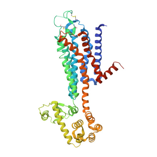
    The adenosine subfamily G protein-coupled receptors A 2A R and A 2B R have been identified as promising cancer immunotherapy candidates. One of the A 2A R/A 2B R dual antagonists, AB928, has progressed to a phase II clinical trial to treat rectal cancer. However, the precise mechanism underlying its dual-antagonistic properties remains elusive. Herein, we report crystal structures of the A 2A R complexed with AB928 and a selective A 2A R antagonist 2-118. The structures revealed a common binding mode on A 2A R, wherein the ligands established extensive interactions with residues from the orthosteric and secondary pockets. In contrast, the cAMP assay and A 2A R and A 2B R molecular dynamics simulations indicated that the ligands adopted distinct binding modes on A 2B R. Detailed analysis of their chemical structures suggested that AB928 readily adapted to the A 2B R pocket, while 2-118 did not due to intrinsic differences. This disparity potentially accounted for the difference in inhibitory efficacy between A 2B R and A 2A R. This study serves as a valuable structural template for the future development of selective or dual inhibitors targeting A 2A R/A 2B R for cancer therapy.


  • Organizational Affiliation

    Shanghai Key Laboratory of Regulatory Biology, Institute of Biomedical Sciences and School of Life Sciences, East China Normal University, Shanghai, 200241, China.


Macromolecules
Find similar proteins by:  (by identity cutoff)  |  3D Structure
Entity ID: 1
MoleculeChains Sequence LengthOrganismDetailsImage
Adenosine receptor A2a,Endolysin481Homo sapiensTequatrovirus T4Mutation(s): 2 
Gene Names: ADORA2AADORA2eT4Tp126
EC: 3.2.1.17
Membrane Entity: Yes 
UniProt & NIH Common Fund Data Resources
Find proteins for P29274 (Homo sapiens)
Explore P29274 
Go to UniProtKB:  P29274
PHAROS:  P29274
GTEx:  ENSG00000128271 
Find proteins for D9IEF7 (Enterobacteria phage T4)
Explore D9IEF7 
Go to UniProtKB:  D9IEF7
Entity Groups  
Sequence Clusters30% Identity50% Identity70% Identity90% Identity95% Identity100% Identity
UniProt GroupsD9IEF7P29274
Sequence Annotations
Expand
  • Reference Sequence
Small Molecules
Ligands 6 Unique
IDChains Name / Formula / InChI Key2D Diagram3D Interactions
U30 (Subject of Investigation/LOI)
Query on U30

Download Ideal Coordinates CCD File 
U [auth A]3-[2-azanyl-6-[1-[[6-(2-oxidanylpropan-2-yl)pyridin-2-yl]methyl]-1,2,3-triazol-4-yl]pyrimidin-4-yl]-2-methyl-benzenecarbonitrile
C23 H22 N8 O
BUXIAWLTBSXYSW-UHFFFAOYSA-N
CLR
Query on CLR

Download Ideal Coordinates CCD File 
Y [auth A]CHOLESTEROL
C27 H46 O
HVYWMOMLDIMFJA-DPAQBDIFSA-N
OLC
Query on OLC

Download Ideal Coordinates CCD File 
Q [auth A],
V [auth A]
(2R)-2,3-dihydroxypropyl (9Z)-octadec-9-enoate
C21 H40 O4
RZRNAYUHWVFMIP-GDCKJWNLSA-N
OLA
Query on OLA

Download Ideal Coordinates CCD File 
B [auth A]
C [auth A]
D [auth A]
E [auth A]
F [auth A]
B [auth A],
C [auth A],
D [auth A],
E [auth A],
F [auth A],
G [auth A],
H [auth A],
J [auth A],
K [auth A],
L [auth A],
M [auth A],
N [auth A],
O [auth A],
P [auth A],
R [auth A],
S [auth A],
T [auth A],
W [auth A]
OLEIC ACID
C18 H34 O2
ZQPPMHVWECSIRJ-KTKRTIGZSA-N
PEG
Query on PEG

Download Ideal Coordinates CCD File 
X [auth A]DI(HYDROXYETHYL)ETHER
C4 H10 O3
MTHSVFCYNBDYFN-UHFFFAOYSA-N
NA
Query on NA

Download Ideal Coordinates CCD File 
I [auth A]SODIUM ION
Na
FKNQFGJONOIPTF-UHFFFAOYSA-N
Experimental Data & Validation

Experimental Data

  • Method: X-RAY DIFFRACTION
  • Resolution: 2.37 Å
  • R-Value Free: 0.240 
  • R-Value Work: 0.189 
  • R-Value Observed: 0.193 
  • Space Group: P 1 21 1
Unit Cell:
Length ( Å )Angle ( ˚ )
a = 46.87α = 90
b = 76.2β = 101.63
c = 85.66γ = 90
Software Package:
Software NamePurpose
Cootmodel building
PHENIXrefinement
Cootmodel building
Cootmodel building
PHASERphasing
XDSdata scaling
XDSdata reduction

Structure Validation

View Full Validation Report


Ligand Structure Quality Assessment 


Entry History & Funding Information

Deposition Data


Funding OrganizationLocationGrant Number
National Natural Science Foundation of China (NSFC)China32171215

Revision History  (Full details and data files)

  • Version 1.0: 2023-08-16
    Type: Initial release
  • Version 1.1: 2024-02-21
    Changes: Database references