8J2F | pdb_00008j2f

Human neutral shpingomyelinase

  • Classification: MEMBRANE PROTEIN
  • Organism(s): Homo sapiens
  • Expression System: Homo sapiens
  • Mutation(s): No 
  • Membrane Protein: Yes  PDBTM

  • Deposited: 2023-04-14 Released: 2023-12-06 
  • Deposition Author(s): Zhang, S.S.
  • Funding Organization(s): National Natural Science Foundation of China (NSFC)

Experimental Data Snapshot

  • Method: ELECTRON MICROSCOPY
  • Resolution: 3.07 Å
  • Aggregation State: PARTICLE 
  • Reconstruction Method: SINGLE PARTICLE 

wwPDB Validation 3D Report Full Report

Validation slider image for 8J2F

This is version 1.1 of the entry. See complete history

Literature

Molecular basis for the catalytic mechanism of human neutral sphingomyelinases 1 (hSMPD2).

Yi, J.Qi, B.Yin, J.Li, R.Chen, X.Hu, J.Li, G.Zhang, S.Zhang, Y.Yang, M.

(2023) Nat Commun 14: 7755-7755

  • DOI: https://doi.org/10.1038/s41467-023-43580-w
  • Primary Citation Related Structures: 
    8J2F

  • PubMed Abstract: 

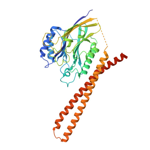
    Enzymatic breakdown of sphingomyelin by sphingomyelinase (SMase) is the main source of the membrane lipids, ceramides, which are involved in many cellular physiological processes. However, the full-length structure of human neutral SMase has not been resolved; therefore, its catalytic mechanism remains unknown. Here, we resolve the structure of human full-length neutral SMase, sphingomyelinase 1 (SMPD2), which reveals that C-terminal transmembrane helices contribute to dimeric architecture of hSMPD2 and that D111 - K116 loop domain is essential for substrate hydrolysis. Coupled with molecular docking, we clarify the binding pose of sphingomyelin, and site-directed mutagenesis further confirms key residues responsible for sphingomyelin binding. Hybrid quantum mechanics/molecular mechanics (QM/MM) molecular dynamic (MD) simulations are utilized to elaborate the catalysis of hSMPD2 with the reported in vitro substrates, sphingomyelin and lyso-platelet activating fator (lyso-PAF). Our study provides mechanistic details that enhance our knowledge of lipid metabolism and may lead to an improved understanding of ceramide in disease and in cancer treatment.


  • Organizational Affiliation
    • Ministry of Education Key Laboratory of Protein Science, Tsinghua-Peking Joint Center for Life Sciences, Beijing Advanced Innovation Center for Structural Biology, School of Life Sciences, Tsinghua University, Beijing, 100084, China.

Macromolecule Content 

  • Total Structure Weight: 96.05 kDa 
  • Atom Count: 6,164 
  • Modeled Residue Count: 768 
  • Deposited Residue Count: 846 
  • Unique protein chains: 1

Macromolecules

Find similar proteins by:|  3D Structure
Entity ID: 1
MoleculeChains  Sequence LengthOrganismDetailsImage
Sphingomyelin phosphodiesterase 2
A, B
423Homo sapiensMutation(s): 0 
Gene Names: SMPD2
EC: 3.1.4.12
Membrane Entity: Yes 
UniProt & NIH Common Fund Data Resources
Find proteins for O60906 (Homo sapiens)
Explore O60906 
Go to UniProtKB:  O60906
PHAROS:  O60906
GTEx:  ENSG00000135587 
Entity Groups
Sequence Clusters30% Identity50% Identity70% Identity90% Identity95% Identity100% Identity
UniProt GroupO60906
Sequence Annotations
Expand
Reference Sequence

Experimental Data & Validation

Experimental Data

  • Method: ELECTRON MICROSCOPY
  • Resolution: 3.07 Å
  • Aggregation State: PARTICLE 
  • Reconstruction Method: SINGLE PARTICLE 

Structure Validation

View Full Validation Report


Entry History 

& Funding Information

Deposition Data

  • Released Date: 2023-12-06 
  • Deposition Author(s): Zhang, S.S.

Funding OrganizationLocationGrant Number
National Natural Science Foundation of China (NSFC)China32030056

Revision History  (Full details and data files)

  • Version 1.0: 2023-12-06
    Type: Initial release
  • Version 1.1: 2024-05-01
    Changes: Database references