8HES | pdb_00008hes

Crystal structure of SARS-CoV-2 RBD and NIV-10 complex


Experimental Data Snapshot

  • Method: X-RAY DIFFRACTION
  • Resolution: 2.20 Å
  • R-Value Free: 
    0.241 (Depositor), 0.240 (DCC) 
  • R-Value Work: 
    0.196 (Depositor), 0.196 (DCC) 
  • R-Value Observed: 
    0.198 (Depositor) 

Starting Model: experimental
View more details

wwPDB Validation 3D Report Full Report

Validation slider image for 8HES

Ligand Structure Quality Assessment 


This is version 1.2 of the entry. See complete history

Literature

Structural delineation and computational design of SARS-CoV-2-neutralizing antibodies against Omicron subvariants.

Moriyama, S.Anraku, Y.Taminishi, S.Adachi, Y.Kuroda, D.Kita, S.Higuchi, Y.Kirita, Y.Kotaki, R.Tonouchi, K.Yumoto, K.Suzuki, T.Someya, T.Fukuhara, H.Kuroda, Y.Yamamoto, T.Onodera, T.Fukushi, S.Maeda, K.Nakamura-Uchiyama, F.Hashiguchi, T.Hoshino, A.Maenaka, K.Takahashi, Y.

(2023) Nat Commun 14: 4198-4198

  • DOI: https://doi.org/10.1038/s41467-023-39890-8
  • Primary Citation Related Structures: 
    7YH6, 7YH7, 8HES, 8HGL, 8HGM

  • PubMed Abstract: 

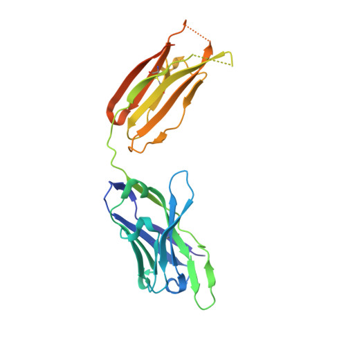
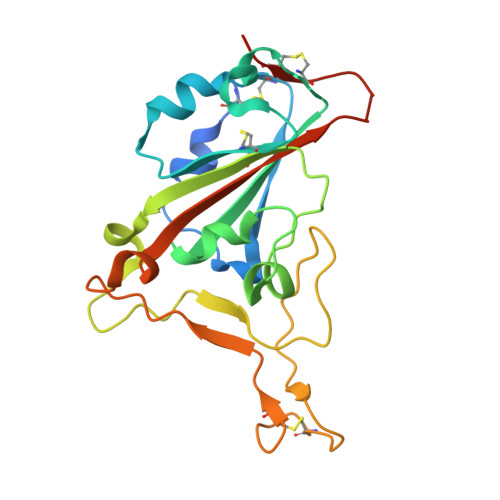
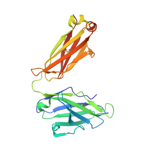
    SARS-CoV-2 Omicron subvariants have evolved to evade receptor-binding site (RBS) antibodies that exist in diverse individuals as public antibody clones. We rationally selected RBS antibodies resilient to mutations in emerging Omicron subvariants. Y489 was identified as a site of virus vulnerability and a common footprint of broadly neutralizing antibodies against the subvariants. Multiple Y489-binding antibodies were encoded by public clonotypes and additionally recognized F486, potentially accounting for the emergence of Omicron subvariants harboring the F486V mutation. However, a subclass of antibodies broadly neutralized BA.4/BA.5 variants via hydrophobic binding sites of rare clonotypes along with high mutation-resilience under escape mutation screening. A computationally designed antibody based on one of the Y489-binding antibodies, NIV-10/FD03, was able to bind XBB with any 486 mutation and neutralized XBB.1.5. The structural basis for the mutation-resilience of this Y489-binding antibody group may provide important insights into the design of therapeutics resistant to viral escape.


  • Organizational Affiliation
    • Research Center for Drug and Vaccine Development, National Institute of Infectious Diseases; Shinjuku-ku, Tokyo, 162-8640, Japan. sayamrym@niid.go.jp.

Macromolecule Content 

  • Total Structure Weight: 74.15 kDa 
  • Atom Count: 4,991 
  • Modeled Residue Count: 621 
  • Deposited Residue Count: 681 
  • Unique protein chains: 3

Macromolecules

Find similar proteins by:|  3D Structure
Entity ID: 1
MoleculeChains  Sequence LengthOrganismDetailsImage
NIV-10 Fab H-chainA [auth H]249Homo sapiensMutation(s): 0 
Find similar proteins by:|  3D Structure
Entity ID: 2
MoleculeChains  Sequence LengthOrganismDetailsImage
Spike protein S1B [auth C]215Severe acute respiratory syndrome coronavirus 2Mutation(s): 0 
UniProt
Find proteins for P0DTC2 (Severe acute respiratory syndrome coronavirus 2)
Explore P0DTC2 
Go to UniProtKB:  P0DTC2
Entity Groups
Sequence Clusters30% Identity50% Identity70% Identity90% Identity95% Identity100% Identity
UniProt GroupP0DTC2
Glycosylation
Glycosylation Sites: 1Go to GlyGen: P0DTC2-1
Sequence Annotations
Expand
Reference Sequence
Find similar proteins by:|  3D Structure
Entity ID: 3
MoleculeChains  Sequence LengthOrganismDetailsImage
NIV-10 Fab L-chainC [auth L]217Homo sapiensMutation(s): 0 

Small Molecules

Ligands 1 Unique
IDChains Name / Formula / InChI Key2D Diagram3D Interactions
NAG

Query on NAG


Download:Ideal Coordinates CCD File
D [auth C]2-acetamido-2-deoxy-beta-D-glucopyranose
C8 H15 N O6
OVRNDRQMDRJTHS-FMDGEEDCSA-N

Experimental Data & Validation

Experimental Data

  • Method: X-RAY DIFFRACTION
  • Resolution: 2.20 Å
  • R-Value Free:  0.241 (Depositor), 0.240 (DCC) 
  • R-Value Work:  0.196 (Depositor), 0.196 (DCC) 
  • R-Value Observed: 0.198 (Depositor) 
Space Group: P 21 21 21
Unit Cell:
Length ( Å )Angle ( ˚ )
a = 53.147α = 90
b = 101.113β = 90
c = 134.265γ = 90
Software Package:
Software NamePurpose
PHENIXrefinement
XDSdata reduction
Aimlessdata scaling
PHASERphasing

Structure Validation

View Full Validation Report


Ligand Structure Quality Assessment 


Entry History 

& Funding Information

Deposition Data


Funding OrganizationLocationGrant Number
Japan Agency for Medical Research and Development (AMED)JapanJP20fk0108516
Japan Agency for Medical Research and Development (AMED)JapanJP 21fk0108465
Japan Agency for Medical Research and Development (AMED)JapanJP20fk0108298
Japan Agency for Medical Research and Development (AMED)JapanJP20fk0108534
Japan Agency for Medical Research and Development (AMED)JapanJP21fk0108534
Japan Agency for Medical Research and Development (AMED)JapanJP19fk0108104
Japan Agency for Medical Research and Development (AMED)JapanJP20fk0108104
Japan Agency for Medical Research and Development (AMED)JapanJP22ama121037
Japan Society for the Promotion of Science (JSPS)JapanJP20H05873
Japan Society for the Promotion of Science (JSPS)JapanJP20H05773

Revision History  (Full details and data files)

  • Version 1.0: 2023-11-08
    Type: Initial release
  • Version 1.1: 2024-10-09
    Changes: Structure summary
  • Version 1.2: 2026-03-04
    Changes: Refinement description