8H3S

Substrate-bound EP, polyA model


Experimental Data Snapshot

  • Method: ELECTRON MICROSCOPY
  • Resolution: 4.90 Å
  • Aggregation State: PARTICLE 
  • Reconstruction Method: SINGLE PARTICLE 

wwPDB Validation   3D Report Full Report


This is version 1.0 of the entry. See complete history


Literature

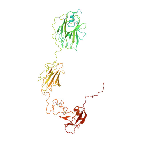
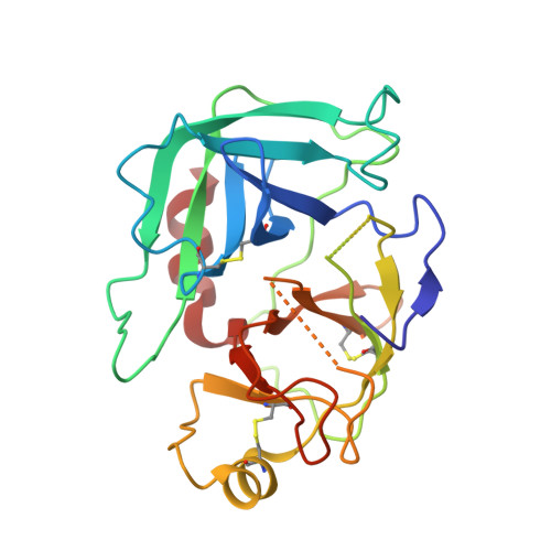
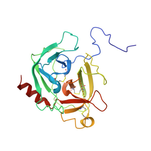
Cryo-EM structures reveal the activation and substrate recognition mechanism of human enteropeptidase.

Yang, X.Ding, Z.Peng, L.Song, Q.Zhang, D.Cui, F.Xia, C.Li, K.Yin, H.Li, S.Li, Z.Huang, H.

(2022) Nat Commun 13: 6955-6955

  • DOI: https://doi.org/10.1038/s41467-022-34364-9
  • Primary Citation of Related Structures:  
    7WQW, 7WQX, 7WQZ, 7WR7, 8H3S, 8H3U

  • PubMed Abstract: 

    Enteropeptidase (EP) initiates intestinal digestion by proteolytically processing trypsinogen, generating catalytically active trypsin. EP dysfunction causes a series of pancreatic diseases including acute necrotizing pancreatitis. However, the molecular mechanisms of EP activation and substrate recognition remain elusive, due to the lack of structural information on the EP heavy chain. Here, we report cryo-EM structures of human EP in inactive, active, and substrate-bound states at resolutions from 2.7 to 4.9 Å. The EP heavy chain was observed to clamp the light chain with CUB2 domain for substrate recognition. The EP light chain N-terminus induced a rearrangement of surface-loops from inactive to active conformations, resulting in activated EP. The heavy chain then served as a hinge for light-chain conformational changes to recruit and subsequently cleave substrate. Our study provides structural insights into rearrangements of EP surface-loops and heavy chain dynamics in the EP catalytic cycle, advancing our understanding of EP-associated pancreatitis.


  • Organizational Affiliation

    Department of Gastroenterology, Changhai Hospital, Navy/Second Military Medical University, Shanghai, China.


Macromolecules
Find similar proteins by:  (by identity cutoff)  |  3D Structure
Entity ID: 1
MoleculeChains Sequence LengthOrganismDetailsImage
Enteropeptidase non-catalytic heavy chain602Homo sapiensMutation(s): 0 
Gene Names: TMPRSS15ENTKPRSS7
UniProt & NIH Common Fund Data Resources
Find proteins for P98073 (Homo sapiens)
Explore P98073 
Go to UniProtKB:  P98073
PHAROS:  P98073
GTEx:  ENSG00000154646 
Entity Groups  
Sequence Clusters30% Identity50% Identity70% Identity90% Identity95% Identity100% Identity
UniProt GroupP98073
Sequence Annotations
Expand
  • Reference Sequence
Find similar proteins by:  (by identity cutoff)  |  3D Structure
Entity ID: 2
MoleculeChains Sequence LengthOrganismDetailsImage
Enteropeptidase catalytic light chain235Homo sapiensMutation(s): 3 
Gene Names: TMPRSS15ENTKPRSS7
UniProt & NIH Common Fund Data Resources
Find proteins for P98073 (Homo sapiens)
Explore P98073 
Go to UniProtKB:  P98073
PHAROS:  P98073
GTEx:  ENSG00000154646 
Entity Groups  
Sequence Clusters30% Identity50% Identity70% Identity90% Identity95% Identity100% Identity
UniProt GroupP98073
Sequence Annotations
Expand
  • Reference Sequence
Find similar proteins by:  (by identity cutoff)  |  3D Structure
Entity ID: 3
MoleculeChains Sequence LengthOrganismDetailsImage
Serine protease 1232Homo sapiensMutation(s): 0 
Gene Names: PRSS1TRP1TRY1TRYP1
EC: 3.4.21.4
UniProt & NIH Common Fund Data Resources
Find proteins for P07477 (Homo sapiens)
Explore P07477 
Go to UniProtKB:  P07477
PHAROS:  P07477
GTEx:  ENSG00000204983 
Entity Groups  
Sequence Clusters30% Identity50% Identity70% Identity90% Identity95% Identity100% Identity
UniProt GroupP07477
Sequence Annotations
Expand
  • Reference Sequence
Experimental Data & Validation

Experimental Data

  • Method: ELECTRON MICROSCOPY
  • Resolution: 4.90 Å
  • Aggregation State: PARTICLE 
  • Reconstruction Method: SINGLE PARTICLE 

Structure Validation

View Full Validation Report


Entry History & Funding Information

Deposition Data


Funding OrganizationLocationGrant Number
Not funded--

Revision History  (Full details and data files)

  • Version 1.0: 2022-11-23
    Type: Initial release