8G8W

Molecular mechanism of nucleotide inhibition of human uncoupling protein 1


Experimental Data Snapshot

  • Method: ELECTRON MICROSCOPY
  • Resolution: 3.80 Å
  • Aggregation State: PARTICLE 
  • Reconstruction Method: SINGLE PARTICLE 

wwPDB Validation   3D Report Full Report


This is version 1.0 of the entry. See complete history


Literature

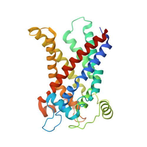
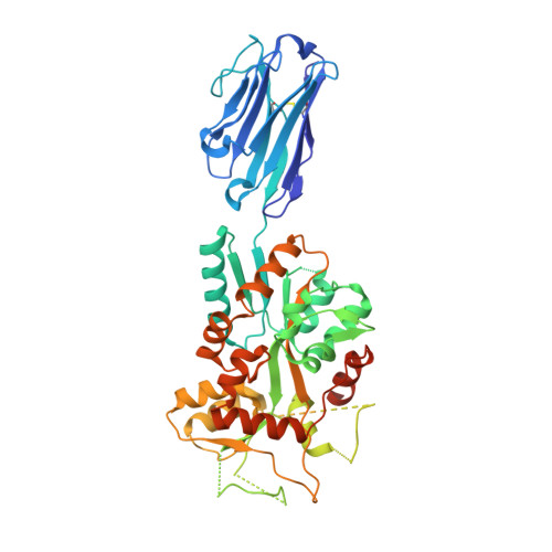
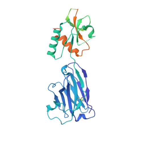
Structural basis of purine nucleotide inhibition of human uncoupling protein 1.

Jones, S.A.Gogoi, P.Ruprecht, J.J.King, M.S.Lee, Y.Zogg, T.Pardon, E.Chand, D.Steimle, S.Copeman, D.M.Cotrim, C.A.Steyaert, J.Crichton, P.G.Moiseenkova-Bell, V.Kunji, E.R.S.

(2023) Sci Adv 9: eadh4251-eadh4251

  • DOI: https://doi.org/10.1126/sciadv.adh4251
  • Primary Citation of Related Structures:  
    8G8W

  • PubMed Abstract: 

    Mitochondrial uncoupling protein 1 (UCP1) gives brown adipose tissue of mammals its specialized ability to burn calories as heat for thermoregulation. When activated by fatty acids, UCP1 catalyzes the leak of protons across the mitochondrial inner membrane, short-circuiting the mitochondrion to generate heat, bypassing ATP synthesis. In contrast, purine nucleotides bind and inhibit UCP1, regulating proton leak by a molecular mechanism that is unclear. We present the cryo-electron microscopy structure of the GTP-inhibited state of UCP1, which is consistent with its nonconducting state. The purine nucleotide cross-links the transmembrane helices of UCP1 with an extensive interaction network. Our results provide a structural basis for understanding the specificity and pH dependency of the regulatory mechanism. UCP1 has retained all of the key functional and structural features required for a mitochondrial carrier-like transport mechanism. The analysis shows that inhibitor binding prevents the conformational changes that UCP1 uses to facilitate proton leak.


  • Organizational Affiliation

    MRC Mitochondrial Biology Unit, University of Cambridge, Cambridge Biomedical Campus, Keith Peters Building, Cambridge CB2 0XY, UK.


Macromolecules
Find similar proteins by:  (by identity cutoff)  |  3D Structure
Entity ID: 1
MoleculeChains Sequence LengthOrganismDetailsImage
Mitochondrial brown fat uncoupling protein 1310Homo sapiensMutation(s): 0 
Gene Names: UCP1SLC25A7UCP
Membrane Entity: Yes 
UniProt & NIH Common Fund Data Resources
Find proteins for P25874 (Homo sapiens)
Explore P25874 
Go to UniProtKB:  P25874
PHAROS:  P25874
GTEx:  ENSG00000109424 
Entity Groups  
Sequence Clusters30% Identity50% Identity70% Identity90% Identity95% Identity100% Identity
UniProt GroupP25874
Sequence Annotations
Expand
  • Reference Sequence
Find similar proteins by:  (by identity cutoff)  |  3D Structure
Entity ID: 2
MoleculeChains Sequence LengthOrganismDetailsImage
Pro-macrobody 71, Maltose/maltodextrin-binding periplasmic protein chimera490synthetic constructEscherichia coli K-12
This entity is chimeric
Mutation(s): 0 
Membrane Entity: Yes 
UniProt
Find proteins for P0AEX9 (Escherichia coli (strain K12))
Explore P0AEX9 
Go to UniProtKB:  P0AEX9
Entity Groups  
Sequence Clusters30% Identity50% Identity70% Identity90% Identity95% Identity100% Identity
UniProt GroupP0AEX9
Sequence Annotations
Expand
  • Reference Sequence
Find similar proteins by:  (by identity cutoff)  |  3D Structure
Entity ID: 3
MoleculeChains Sequence LengthOrganismDetailsImage
Pro-Macrobody 65, Maltose/maltodextrin-binding periplasmic protein chimera494synthetic constructEscherichia coli K-12
This entity is chimeric
Mutation(s): 0 
Membrane Entity: Yes 
UniProt
Find proteins for P0AEX9 (Escherichia coli (strain K12))
Explore P0AEX9 
Go to UniProtKB:  P0AEX9
Entity Groups  
Sequence Clusters30% Identity50% Identity70% Identity90% Identity95% Identity100% Identity
UniProt GroupP0AEX9
Sequence Annotations
Expand
  • Reference Sequence
Oligosaccharides

Help

Entity ID: 4
MoleculeChains Length2D Diagram Glycosylation3D Interactions
alpha-D-glucopyranose-(1-4)-alpha-D-glucopyranose
D
2N/A
Glycosylation Resources
GlyTouCan:  G07411ON
GlyCosmos:  G07411ON
Experimental Data & Validation

Experimental Data

  • Method: ELECTRON MICROSCOPY
  • Resolution: 3.80 Å
  • Aggregation State: PARTICLE 
  • Reconstruction Method: SINGLE PARTICLE 

Structure Validation

View Full Validation Report


Entry History & Funding Information

Deposition Data


Funding OrganizationLocationGrant Number
National Institutes of Health/National Institute of General Medical Sciences (NIH/NIGMS)United StatesGM144120
Medical Research Council (MRC, United Kingdom)United KingdomMC_UU_00028/2
Medical Research Council (MRC, United Kingdom)United KingdomMC_UU_00015/1
Biotechnology and Biological Sciences Research Council (BBSRC)United KingdomBB/S00940X/1
European Union (EU)European UnionInstruct-ERIC/ESFRI
Research Foundation - Flanders (FWO)Belgium--

Revision History  (Full details and data files)

  • Version 1.0: 2023-06-07
    Type: Initial release