8FXT

Escherichia coli periplasmic Glucose-Binding Protein glucose complex: Acrylodan conjugate attached at W183C


Experimental Data Snapshot

  • Method: X-RAY DIFFRACTION
  • Resolution: 1.53 Å
  • R-Value Free: 0.183 
  • R-Value Work: 0.155 
  • R-Value Observed: 0.157 

wwPDB Validation   3D Report Full Report


This is version 1.0 of the entry. See complete history


Literature

Chromophore carbonyl twisting in fluorescent biosensors encodes direct readout of protein conformations with multicolor switching.

Allert, M.J.Kumar, S.Wang, Y.Beese, L.S.Hellinga, H.W.

(2023) Commun Chem 6: 168-168

  • DOI: https://doi.org/10.1038/s42004-023-00982-7
  • Primary Citation of Related Structures:  
    8FXT, 8FXU

  • PubMed Abstract: 

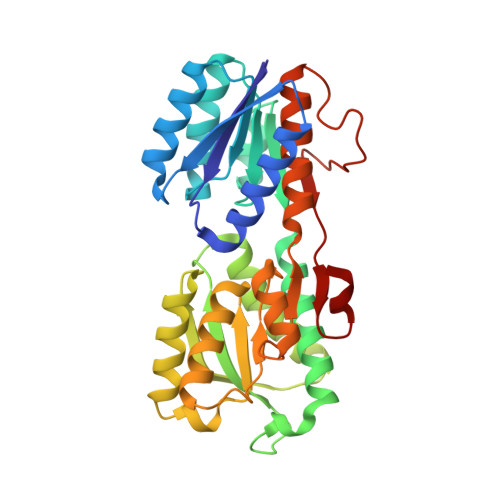
    Fluorescent labeling of proteins is a powerful tool for probing structure-function relationships with many biosensing applications. Structure-based rules for systematically designing fluorescent biosensors require understanding ligand-mediated fluorescent response mechanisms which can be challenging to establish. We installed thiol-reactive derivatives of the naphthalene-based fluorophore Prodan into bacterial periplasmic glucose-binding proteins. Glucose binding elicited paired color exchanges in the excited and ground states of these conjugates. X-ray structures and mutagenesis studies established that glucose-mediated color switching arises from steric interactions that couple protein conformational changes to twisting of the Prodan carbonyl relative to its naphthalene plane. Mutations of residues contacting the carbonyl can optimize color switching by altering fluorophore conformational equilibria in the apo and glucose-bound proteins. A commonly accepted view is that Prodan derivatives report on protein conformations via solvatochromic effects due to changes in the dielectric of their local environment. Here we show that instead Prodan carbonyl twisting controls color switching. These insights enable structure-based biosensor design by coupling ligand-mediated protein conformational changes to internal chromophore twists through specific steric interactions between fluorophore and protein.


  • Organizational Affiliation

    Department of Biochemistry and Molecular Biophysics, Washington University in St. Louis, St. Louis, MO, 63110, USA.


Macromolecules
Find similar proteins by:  (by identity cutoff)  |  3D Structure
Entity ID: 1
MoleculeChains Sequence LengthOrganismDetailsImage
D-galactose/methyl-galactoside binding periplasmic protein MglB305Escherichia coliMutation(s): 1 
Gene Names: 
mglBA5U30_003457A6592_11845A8499_001034A9X72_08630AAG43_000117ACN68_22070ACN81_16630ACU57_09035AM464_02030APX88_04775AT845_003264AW119_06275B6R12_000806B6R15_002974B6R31_001105B6R48_001523B6R87_001412BANRA_02934BANRA_03201BEA19_19205BF481_000566BG944_000404BGM66_000282BHS81_13365BJI68_13425BJJ90_08540BK292_04615BK383_00945BLM69_000640BMC79_002822BMT50_20295BMT91_00320BON92_16595BR158_000766BRV02_000218BSR05_15935BTB68_001398BTQ06_09915BUO55_000779BvCmsKKP061_01094BXT93_06625BZL69_09460C0P57_000336C1Q91_000149C2121_000044C2R31_001598C3F40_26965C5N07_04915C9114_02655CA593_16510CCS08_24120CDL36_00775CDL37_14560CF22_000075CG831_003047CIG67_25325CO706_08460CQ986_001545CQB02_05580CR538_08960CR539_15575CR628_000919CTR35_000577CV83915_04058CWS33_00530CX938_002180CY655_14220D0X26_13110D1912_29380D3C88_28390D3G36_01935D4M65_09620D4N09_08940D4U49_06260D9D43_10510D9E34_15485D9E49_00365D9H94_00195D9J03_10425D9J61_06325DD762_02400DIV22_25860DN627_05125DNQ45_17790DNX30_05415DRW19_12180DS732_16775DTL43_05580DTL90_20525DU321_11675E0I42_04230E3N34_19380E4K51_00995E5H86_03650E6D34_12480EA239_06510EA435_00700EAI46_17175EAN77_00390EAX79_04555EBP16_01130EC95NR1_01244EHD79_01145EIA08_04520EIZ93_07035EKI52_24795EL79_1498ELT17_20450ELT48_05005ELX68_00965ELX76_00300ELX79_03655ELX96_01815ELY41_06365ELY48_03520EN85_000169EPS76_11515ERS085406_00895EWK56_06995ExPECSC038_02176F3N40_00545F7F11_05695F7N46_17820F9413_06505F9461_14760F9B07_08545F9S83_05355FDM60_10915FEJ01_13975FFF58_20910FGG80_11195FHD44_01870FIJ20_04990FJQ40_11760FJQ53_07810FKO60_02375FOI11_002185FOI11_17860FPI65_13600FPS11_10035FV293_14455FVB16_21795FZU14_08145G3565_00740G3V95_00210G4A38_04815G4A47_00180G5603_05160G9448_23120GAI89_01085GAJ12_16505GAJ26_13230GF699_16440GFY34_10720GIB53_03975GJ11_14600GJO56_00690GKF66_07705GKF89_02955GNW61_03440GOP25_08560GP711_03870GP944_07610GP975_08300GQM04_13060GQM13_02295GQM21_09055GQN34_07310GQW07_17680GRC73_02015GRO95_17210GRW05_06490GRW24_11420GSM54_08360GSY44_10065GUC01_05135GUI33_20580H0O51_12230H0O72_00925H6Y26_001607HCQ42_002157HEP30_015805HEP34_000322HHH44_004011HI055_001096HIE29_002648HJQ60_001810HKA49_000580HL563_04405HL601_00440HLV18_10300HLX92_07325HLZ50_13230HMV95_15185HMW38_03860HV109_08050HV146_14830HV209_10285HVV39_02595HVW04_24210HVW43_25785HVY77_09315HVZ71_09125HX136_08220I6H00_01185I6H02_06005IA00_003210IFB95_000112IH772_22875IT029_002351J0541_002029J4S20_001455J5U05_001805JE86ST02C_25870JE86ST05C_26720JFD_00293JNP96_18035NCTC10082_04144NCTC10089_01711NCTC10090_04751NCTC10418_02713NCTC10429_02796NCTC10764_01024NCTC10865_02242NCTC10974_02040NCTC11181_03969NCTC11341_00467NCTC13216_04862NCTC4450_03719NCTC7922_01437NCTC7927_01980NCTC7928_00841NCTC8009_03554NCTC8179_00574NCTC8450_03770NCTC8500_01967NCTC8959_02087NCTC8960_04581NCTC9044_05932NCTC9045_02047NCTC9073_01492NCTC9075_02461NCTC9077_02204NCTC9081_05093NCTC9111_05562NCTC9117_02380NCTC9702_02039NCTC9775_05615NCTC9777_05224ND22_000990RG28_11230SAMEA3472056_04006SAMEA3751407_01797SAMEA3752557_00469SAMEA3753106_00196TUM18780_15170WR15_09685

UniProt
Find proteins for E2QP16 (Escherichia coli)
Explore E2QP16 
Go to UniProtKB:  E2QP16
Entity Groups  
Sequence Clusters30% Identity50% Identity70% Identity90% Identity95% Identity100% Identity
UniProt GroupE2QP16
Sequence Annotations
Expand
  • Reference Sequence
Small Molecules
Ligands 4 Unique
IDChains Name / Formula / InChI Key2D Diagram3D Interactions
YDM (Subject of Investigation/LOI)
Query on YDM

Download Ideal Coordinates CCD File 
B [auth A]1-[6-(dimethylamino)naphthalen-2-yl]propan-1-one
C15 H17 N O
MPPQGYCZBNURDG-UHFFFAOYSA-N
BGC
Query on BGC

Download Ideal Coordinates CCD File 
D [auth A],
E [auth A],
F [auth A],
G [auth A],
H [auth A]
beta-D-glucopyranose
C6 H12 O6
WQZGKKKJIJFFOK-VFUOTHLCSA-N
GLC
Query on GLC

Download Ideal Coordinates CCD File 
C [auth A]alpha-D-glucopyranose
C6 H12 O6
WQZGKKKJIJFFOK-DVKNGEFBSA-N
CA
Query on CA

Download Ideal Coordinates CCD File 
I [auth A]CALCIUM ION
Ca
BHPQYMZQTOCNFJ-UHFFFAOYSA-N
Experimental Data & Validation

Experimental Data

  • Method: X-RAY DIFFRACTION
  • Resolution: 1.53 Å
  • R-Value Free: 0.183 
  • R-Value Work: 0.155 
  • R-Value Observed: 0.157 
  • Space Group: C 1 2 1
Unit Cell:
Length ( Å )Angle ( ˚ )
a = 119.587α = 90
b = 36.533β = 124.13
c = 79.905γ = 90
Software Package:
Software NamePurpose
PHENIXrefinement
HKL-2000data reduction
HKL-2000data scaling
PHENIXphasing

Structure Validation

View Full Validation Report


Entry History & Funding Information

Deposition Data


Funding OrganizationLocationGrant Number
Becton-Dickinson and CompanyUnited States--

Revision History  (Full details and data files)

  • Version 1.0: 2023-08-30
    Type: Initial release