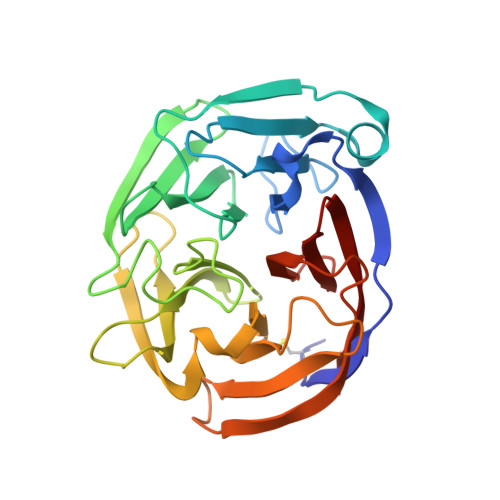
Competition between inside-out unfolding and pathogenic aggregation in an amyloid-forming beta-propeller.
Saccuzzo, E.G., Mebrat, M.D., Scelsi, H.F., Kim, M., Ma, M.T., Su, X., Hill, S.E., Rheaume, E., Li, R., Torres, M.P., Gumbart, J.C., Van Horn, W.D., Lieberman, R.L.(2024) Nat Commun 15: 155-155
- PubMed: 38168102
- DOI: https://doi.org/10.1038/s41467-023-44479-2
- Primary Citation of Related Structures:
8FRR - PubMed Abstract:
Studies of folded-to-misfolded transitions using model protein systems reveal a range of unfolding needed for exposure of amyloid-prone regions for subsequent fibrillization. Here, we probe the relationship between unfolding and aggregation for glaucoma-associated myocilin. Mutations within the olfactomedin domain of myocilin (OLF) cause a gain-of-function, namely cytotoxic intracellular aggregation, which hastens disease progression. Aggregation by wild-type OLF (OLF WT ) competes with its chemical unfolding, but only below the threshold where OLF loses tertiary structure. Representative moderate (OLF D380A ) and severe (OLF I499F ) disease variants aggregate differently, with rates comparable to OLF WT in initial stages of unfolding, and variants adopt distinct partially folded structures seen along the OLF WT urea-unfolding pathway. Whether initiated with mutation or chemical perturbation, unfolding propagates outward to the propeller surface. In sum, for this large protein prone to amyloid formation, the requirement for a conformational change to promote amyloid fibrillization leads to direct competition between unfolding and aggregation.
- School of Chemistry & Biochemistry, Georgia Institute of Technology, Atlanta, USA.
Organizational Affiliation:


