8FRR | pdb_00008frr

Wild-type myocilin olfactomedin domain


Experimental Data Snapshot

  • Method: X-RAY DIFFRACTION
  • Resolution: 1.27 Å
  • R-Value Free: 
    0.155 (Depositor), 0.156 (DCC) 
  • R-Value Work: 
    0.140 (Depositor), 0.140 (DCC) 
  • R-Value Observed: 
    0.140 (Depositor) 

Starting Model: experimental
View more details

wwPDB Validation 3D Report Full Report

Validation slider image for 8FRR

Ligand Structure Quality Assessment 


This is version 1.1 of the entry. See complete history

Literature

Competition between inside-out unfolding and pathogenic aggregation in an amyloid-forming beta-propeller.

Saccuzzo, E.G.Mebrat, M.D.Scelsi, H.F.Kim, M.Ma, M.T.Su, X.Hill, S.E.Rheaume, E.Li, R.Torres, M.P.Gumbart, J.C.Van Horn, W.D.Lieberman, R.L.

(2024) Nat Commun 15: 155-155

  • DOI: https://doi.org/10.1038/s41467-023-44479-2
  • Primary Citation Related Structures: 
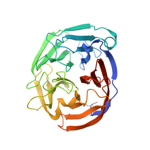
    8FRR

  • PubMed Abstract: 

    Studies of folded-to-misfolded transitions using model protein systems reveal a range of unfolding needed for exposure of amyloid-prone regions for subsequent fibrillization. Here, we probe the relationship between unfolding and aggregation for glaucoma-associated myocilin. Mutations within the olfactomedin domain of myocilin (OLF) cause a gain-of-function, namely cytotoxic intracellular aggregation, which hastens disease progression. Aggregation by wild-type OLF (OLF WT ) competes with its chemical unfolding, but only below the threshold where OLF loses tertiary structure. Representative moderate (OLF D380A ) and severe (OLF I499F ) disease variants aggregate differently, with rates comparable to OLF WT in initial stages of unfolding, and variants adopt distinct partially folded structures seen along the OLF WT urea-unfolding pathway. Whether initiated with mutation or chemical perturbation, unfolding propagates outward to the propeller surface. In sum, for this large protein prone to amyloid formation, the requirement for a conformational change to promote amyloid fibrillization leads to direct competition between unfolding and aggregation.


  • Organizational Affiliation
    • School of Chemistry & Biochemistry, Georgia Institute of Technology, Atlanta, USA.

Macromolecule Content 

  • Total Structure Weight: 29.41 kDa 
  • Atom Count: 2,544 
  • Modeled Residue Count: 259 
  • Deposited Residue Count: 259 
  • Unique protein chains: 1

Macromolecules

Find similar proteins by:|  3D Structure
Entity ID: 1
MoleculeChains  Sequence LengthOrganismDetailsImage
Myocilin, C-terminal fragment259Homo sapiensMutation(s): 0 
Gene Names: MYOCGLC1ATIGR
UniProt & NIH Common Fund Data Resources
Find proteins for Q99972 (Homo sapiens)
Explore Q99972 
Go to UniProtKB:  Q99972
PHAROS:  Q99972
GTEx:  ENSG00000034971 
Entity Groups
Sequence Clusters30% Identity50% Identity70% Identity90% Identity95% Identity100% Identity
UniProt GroupQ99972
Sequence Annotations
Expand
Reference Sequence

Experimental Data & Validation

Experimental Data

  • Method: X-RAY DIFFRACTION
  • Resolution: 1.27 Å
  • R-Value Free:  0.155 (Depositor), 0.156 (DCC) 
  • R-Value Work:  0.140 (Depositor), 0.140 (DCC) 
  • R-Value Observed: 0.140 (Depositor) 
Space Group: P 1 21 1
Unit Cell:
Length ( Å )Angle ( ˚ )
a = 49.468α = 90
b = 50.527β = 96.25
c = 50.873γ = 90
Software Package:
Software NamePurpose
HKL-2000data scaling
PHENIXrefinement
PDB_EXTRACTdata extraction
PHENIXphasing

Structure Validation

View Full Validation Report


Ligand Structure Quality Assessment 


Entry History 

& Funding Information

Deposition Data


Funding OrganizationLocationGrant Number
National Institutes of Health/National Eye Institute (NIH/NEI)United States10R01EY021205

Revision History  (Full details and data files)

  • Version 1.0: 2024-01-17
    Type: Initial release
  • Version 1.1: 2024-10-09
    Changes: Structure summary