8FHD

Cryo-EM structure of human voltage-gated sodium channel Nav1.6


Experimental Data Snapshot

  • Method: ELECTRON MICROSCOPY
  • Resolution: 3.10 Å
  • Aggregation State: PARTICLE 
  • Reconstruction Method: SINGLE PARTICLE 

wwPDB Validation   3D Report Full Report


This is version 1.0 of the entry. See complete history


Literature

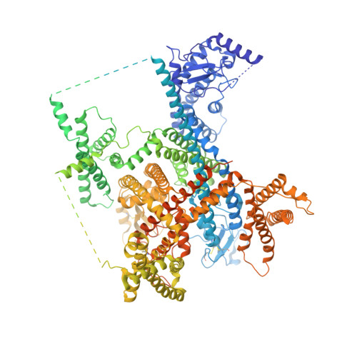
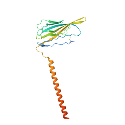
Cryo-EM structure of human voltage-gated sodium channel Na v 1.6.

Fan, X.Huang, J.Jin, X.Yan, N.

(2023) Proc Natl Acad Sci U S A 120: e2220578120-e2220578120

  • DOI: https://doi.org/10.1073/pnas.2220578120
  • Primary Citation of Related Structures:  
    8FHD

  • PubMed Abstract: 

    Voltage-gated sodium channel Na v 1.6 plays a crucial role in neuronal firing in the central nervous system (CNS). Aberrant function of Na v 1.6 may lead to epilepsy and other neurological disorders. Specific inhibitors of Na v 1.6 thus have therapeutic potentials. Here we present the cryo-EM structure of human Na v 1.6 in the presence of auxiliary subunits β1 and fibroblast growth factor homologous factor 2B (FHF2B) at an overall resolution of 3.1 Å. The overall structure represents an inactivated state with closed pore domain (PD) and all "up" voltage-sensing domains. A conserved carbohydrate-aromatic interaction involving Trp302 and Asn326, together with the β1 subunit, stabilizes the extracellular loop in repeat I. Apart from regular lipids that are resolved in the EM map, an unprecedented Y-shaped density that belongs to an unidentified molecule binds to the PD, revealing a potential site for developing Na v 1.6-specific blockers. Structural mapping of disease-related Na v 1.6 mutations provides insights into their pathogenic mechanism.


  • Organizational Affiliation

    Department of Molecular Biology, Princeton University, Princeton, NJ 08544.


Macromolecules
Find similar proteins by:  (by identity cutoff)  |  3D Structure
Entity ID: 1
MoleculeChains Sequence LengthOrganismDetailsImage
Sodium channel protein type 8 subunit alpha1,980Homo sapiensMutation(s): 0 
Gene Names: SCN8AMED
Membrane Entity: Yes 
UniProt & NIH Common Fund Data Resources
Find proteins for Q9UQD0 (Homo sapiens)
Explore Q9UQD0 
Go to UniProtKB:  Q9UQD0
PHAROS:  Q9UQD0
GTEx:  ENSG00000196876 
Entity Groups  
Sequence Clusters30% Identity50% Identity70% Identity90% Identity95% Identity100% Identity
UniProt GroupQ9UQD0
Sequence Annotations
Expand
  • Reference Sequence
Find similar proteins by:  (by identity cutoff)  |  3D Structure
Entity ID: 2
MoleculeChains Sequence LengthOrganismDetailsImage
Sodium channel subunit beta-1B [auth C]218Homo sapiensMutation(s): 0 
Gene Names: SCN1B
Membrane Entity: Yes 
UniProt & NIH Common Fund Data Resources
Find proteins for Q07699 (Homo sapiens)
Explore Q07699 
Go to UniProtKB:  Q07699
PHAROS:  Q07699
GTEx:  ENSG00000105711 
Entity Groups  
Sequence Clusters30% Identity50% Identity70% Identity90% Identity95% Identity100% Identity
UniProt GroupQ07699
Sequence Annotations
Expand
  • Reference Sequence
Oligosaccharides

Help

Entity ID: 3
MoleculeChains Length2D Diagram Glycosylation3D Interactions
alpha-D-mannopyranose-(1-2)-alpha-D-mannopyranose-(1-6)-[alpha-D-mannopyranose-(1-3)]alpha-D-mannopyranose-(1-6)-[alpha-D-mannopyranose-(1-2)-alpha-D-mannopyranose-(1-3)]beta-D-mannopyranose-(1-4)-2-acetamido-2-deoxy-beta-D-glucopyranose-(1-4)-2-acetamido-2-deoxy-beta-D-glucopyranoseC [auth D]9N/AN-Glycosylation
Glycosylation Resources
GlyTouCan:  G83161QT
GlyCosmos:  G83161QT
GlyGen:  G83161QT
Entity ID: 4
MoleculeChains Length2D Diagram Glycosylation3D Interactions
2-acetamido-2-deoxy-beta-D-glucopyranose-(1-4)-2-acetamido-2-deoxy-beta-D-glucopyranoseD [auth F]2N/AN-Glycosylation
Glycosylation Resources
GlyTouCan:  G42666HT
GlyCosmos:  G42666HT
GlyGen:  G42666HT
Small Molecules
Ligands 8 Unique
IDChains Name / Formula / InChI Key2D Diagram3D Interactions
P5S
Query on P5S

Download Ideal Coordinates CCD File 
Q [auth A],
W [auth A]
O-[(R)-{[(2R)-2,3-bis(octadecanoyloxy)propyl]oxy}(hydroxy)phosphoryl]-L-serine
C42 H82 N O10 P
TZCPCKNHXULUIY-RGULYWFUSA-N
PCW
Query on PCW

Download Ideal Coordinates CCD File 
M [auth A],
P [auth A],
R [auth A]
1,2-DIOLEOYL-SN-GLYCERO-3-PHOSPHOCHOLINE
C44 H85 N O8 P
SNKAWJBJQDLSFF-NVKMUCNASA-O
P3X
Query on P3X

Download Ideal Coordinates CCD File 
Y [auth A](5E,17R,20S)-23-amino-20-hydroxy-14,20-dioxo-15,19,21-trioxa-20lambda~5~-phosphatricos-5-en-17-yl hexadecanoate
C35 H68 N O8 P
IXIBEFBSXYIWMP-VCRBCDDQSA-N
9Z9
Query on 9Z9

Download Ideal Coordinates CCD File 
X [auth A](3beta,14beta,17beta,25R)-3-[4-methoxy-3-(methoxymethyl)butoxy]spirost-5-en
C34 H56 O5
CEEBZAXXSRFQIC-GZSGZGDASA-N
LPE
Query on LPE

Download Ideal Coordinates CCD File 
L [auth A],
N [auth A],
O [auth A],
S [auth A],
T [auth A]
1-O-OCTADECYL-SN-GLYCERO-3-PHOSPHOCHOLINE
C26 H57 N O6 P
XKBJVQHMEXMFDZ-AREMUKBSSA-O
Y01
Query on Y01

Download Ideal Coordinates CCD File 
I [auth A],
K [auth A],
U [auth A],
V [auth A]
CHOLESTEROL HEMISUCCINATE
C31 H50 O4
WLNARFZDISHUGS-MIXBDBMTSA-N
CLR
Query on CLR

Download Ideal Coordinates CCD File 
J [auth A]CHOLESTEROL
C27 H46 O
HVYWMOMLDIMFJA-DPAQBDIFSA-N
NAG
Query on NAG

Download Ideal Coordinates CCD File 
AA [auth C]
BA [auth C]
E [auth A]
F [auth A]
G [auth A]
AA [auth C],
BA [auth C],
E [auth A],
F [auth A],
G [auth A],
H [auth A],
Z [auth C]
2-acetamido-2-deoxy-beta-D-glucopyranose
C8 H15 N O6
OVRNDRQMDRJTHS-FMDGEEDCSA-N
Experimental Data & Validation

Experimental Data

  • Method: ELECTRON MICROSCOPY
  • Resolution: 3.10 Å
  • Aggregation State: PARTICLE 
  • Reconstruction Method: SINGLE PARTICLE 

Structure Validation

View Full Validation Report


Entry History & Funding Information

Deposition Data


Funding OrganizationLocationGrant Number
Human Frontier Science Program (HFSP)FranceLT000754

Revision History  (Full details and data files)

  • Version 1.0: 2023-02-08
    Type: Initial release