Tetramerization is essential for the enzymatic function of the Pseudomonas aeruginosa virulence factor UDP-glucose pyrophosphorylase.
Dirr, L., Cleeves, S., Ramon Roth, I., Li, L., Fiebig, T., Ve, T., Haussler, S., Braun, A., von Itzstein, M., Fuhring, J.I.(2024) mBio 15: e0211423-e0211423
- PubMed: 38470050 Search on PubMedSearch on PubMed Central
- DOI: https://doi.org/10.1128/mbio.02114-23
- Primary Citation Related Structures:
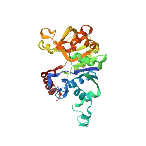
8F73 - PubMed Abstract:
Multidrug-resistant bacteria such as the opportunistic pathogen Pseudomonas aeruginosa , which causes life-threatening infections especially in immunocompromised individuals and cystic fibrosis patients, pose an increasing threat to public health. In the search for new treatment options, P. aeruginosa uridine diphosphate-glucose pyrophosphorylase (PaUGP) has been proposed as a novel drug target because it is required for the biosynthesis of important virulence factors and linked to pathogenicity in animal models. Here, we show that UGP-deficient P. aeruginosa exhibits severely reduced virulence against human lung tissue and cells, emphasizing the enzyme's suitability as a drug target. To establish a basis for the development of selective PaUGP inhibitors, we solved the product-bound crystal structure of tetrameric PaUGP and conducted a comprehensive structure-function analysis, identifying key residues at two different molecular interfaces that are essential for tetramer integrity and catalytic activity and demonstrating that tetramerization is pivotal for PaUGP function. Importantly, we show that part of the PaUGP oligomerization interface is uniquely conserved across bacterial UGPs but does not exist in the human enzyme, therefore representing an allosteric site that may be targeted to selectively inhibit bacterial UGPs.IMPORTANCEInfections with the opportunistic bacterial pathogen Pseudomonas aeruginosa are becoming increasingly difficult to treat due to multidrug resistance. Here, we show that the enzyme uridine diphosphate-glucose pyrophosphorylase (UGP) is involved in P. aeruginosa virulence toward human lung tissue and cells, making it a potential target for the development of new antibacterial drugs. Our exploration of P. aeruginosa (Pa)UGP structure-function relationships reveals that the activity of PaUGP depends on the formation of a tetrameric enzyme complex. We found that a molecular interface involved in tetramer formation is conserved in all bacterial UGPs but not in the human enzyme, and therefore hypothesize that it provides an ideal point of attack to selectively inhibit bacterial UGPs and exploit them as drug targets.
- Institute for Glycomics, Gold Coast Campus, Griffith University, Gold Coast, Queensland, Australia.
Organizational Affiliation: