Structural and functional characterization of the nucleotide-binding domains of ABCA4 and their role in Stargardt disease.
Scortecci, J.F., Garces, F.A., Mahto, J.K., Molday, L.L., Van Petegem, F., Molday, R.S.(2024) J Biol Chem 300: 107666-107666
- PubMed: 39128720 Search on PubMed
- DOI: https://doi.org/10.1016/j.jbc.2024.107666
- Primary Citation Related Structures:
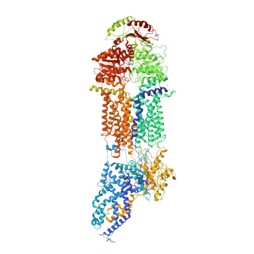
8F5B - PubMed Abstract:
ABCA4 is an ATP-binding cassette (ABC) transporter that prevents the buildup of toxic retinoid compounds by facilitating the transport of N-retinylidene-phosphatidylethanolamine across membranes of rod and cone photoreceptor cells. Over 1500 missense mutations in ABCA4, many in the nucleotide binding domains (NBDs), have been genetically linked to Stargardt disease (STGD1). Here, we show by Cryo-electron microscopy that ABCA4 is converted from an open outward conformation to a closed conformation upon the binding of AMP-PNP. Structural information and biochemical studies were used to further define the role of the NBDs in the functional properties of ABCA4 and the mechanisms by which mutations lead to the loss in activity. We show that ATPase activity in both NBDs is required for the functional activity of ABCA4. Mutations in Walker A asparagine residues cause a severe reduction in substrate-activated ATPase activity due to the loss in polar interactions with residues within the D-loops of the opposing NBD. The structural basis for how disease mutations in other NBD residues including the R1108C, R2077W, R2107H and L2027F affect the structure and function of ABCA4 is described. Collectively, our studies provide insight into the structure and function of ABCA4 and mechanisms underlying STGD1.
- Department of Biochemistry & Molecular Biology, University of British Columbia, Vancouver, B.C. Canada.
Organizational Affiliation: