8E2L

Structure of Lates calcarifer Twinkle helicase with ATP and DNA


Experimental Data Snapshot

  • Method: ELECTRON MICROSCOPY
  • Resolution: 3.51 Å
  • Aggregation State: PARTICLE 
  • Reconstruction Method: SINGLE PARTICLE 

wwPDB Validation   3D Report Full Report


This is version 1.2 of the entry. See complete history


Literature

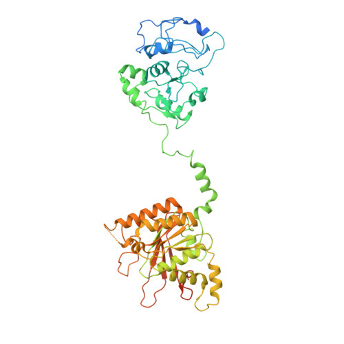
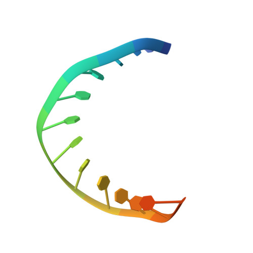
Structural and dynamic basis of DNA capture and translocation by mitochondrial Twinkle helicase.

Li, Z.Kaur, P.Lo, C.Y.Chopra, N.Smith, J.Wang, H.Gao, Y.

(2022) Nucleic Acids Res 50: 11965-11978

  • DOI: https://doi.org/10.1093/nar/gkac1089
  • Primary Citation of Related Structures:  
    8E2L

  • PubMed Abstract: 

    Twinkle is a mitochondrial replicative helicase which can self-load onto and unwind mitochondrial DNA. Nearly 60 mutations on Twinkle have been linked to human mitochondrial diseases. Using cryo-electron microscopy (cryo-EM) and high-speed atomic force microscopy (HS-AFM), we obtained the atomic-resolution structure of a vertebrate Twinkle homolog with DNA and captured in real-time how Twinkle is self-loaded onto DNA. Our data highlight the important role of the non-catalytic N-terminal domain of Twinkle. The N-terminal domain directly contacts the C-terminal helicase domain, and the contact interface is a hotspot for disease-related mutations. Mutations at the interface destabilize Twinkle hexamer and reduce helicase activity. With HS-AFM, we observed that a highly dynamic Twinkle domain, which is likely to be the N-terminal domain, can protrude ∼5 nm to transiently capture nearby DNA and initialize Twinkle loading onto DNA. Moreover, structural analysis and subunit doping experiments suggest that Twinkle hydrolyzes ATP stochastically, which is distinct from related helicases from bacteriophages.


  • Organizational Affiliation

    BioSciences Department, Rice University, Houston, TX 77005, USA.


Macromolecules

Find similar proteins by:  (by identity cutoff)  |  3D Structure
Entity ID: 1
MoleculeChains Sequence LengthOrganismDetailsImage
Twinkle mtDNA helicase542Lates calcariferMutation(s): 1 
UniProt
Find proteins for A0A4W6C5C5 (Lates calcarifer)
Explore A0A4W6C5C5 
Go to UniProtKB:  A0A4W6C5C5
Entity Groups  
Sequence Clusters30% Identity50% Identity70% Identity90% Identity95% Identity100% Identity
UniProt GroupA0A4W6C5C5
Sequence Annotations
Expand
  • Reference Sequence

Find similar nucleic acids by:  Sequence   |   3D Structure  

Entity ID: 2
MoleculeChains LengthOrganismImage
DNA (5'-D(P*TP*TP*TP*TP*TP*TP*TP*TP*TP*TP*TP*T)-3')B [auth M]15synthetic construct
Sequence Annotations
Expand
  • Reference Sequence
Small Molecules
Ligands 2 Unique
IDChains Name / Formula / InChI Key2D Diagram3D Interactions
ATP (Subject of Investigation/LOI)
Query on ATP

Download Ideal Coordinates CCD File 
H [auth C],
J [auth B],
L [auth D],
N [auth E],
P [auth F]
ADENOSINE-5'-TRIPHOSPHATE
C10 H16 N5 O13 P3
ZKHQWZAMYRWXGA-KQYNXXCUSA-N
MG (Subject of Investigation/LOI)
Query on MG

Download Ideal Coordinates CCD File 
I [auth C],
K [auth B],
M [auth D],
O [auth E],
Q [auth F]
MAGNESIUM ION
Mg
JLVVSXFLKOJNIY-UHFFFAOYSA-N
Experimental Data & Validation

Experimental Data

  • Method: ELECTRON MICROSCOPY
  • Resolution: 3.51 Å
  • Aggregation State: PARTICLE 
  • Reconstruction Method: SINGLE PARTICLE 
EM Software:
TaskSoftware PackageVersion
RECONSTRUCTIONcryoSPARC

Structure Validation

View Full Validation Report


Entry History & Funding Information

Deposition Data

  • Released Date: 2022-11-02 
  • Deposition Author(s): Gao, Y., Li, Z.

Funding OrganizationLocationGrant Number
Cancer Prevention and Research Institute of Texas (CPRIT)United StatesRR190046
National Institutes of Health/National Institute of General Medical Sciences (NIH/NIGMS)United StatesGM142722

Revision History  (Full details and data files)

  • Version 1.0: 2022-11-02
    Type: Initial release
  • Version 1.1: 2022-11-30
    Changes: Database references
  • Version 1.2: 2022-12-21
    Changes: Database references