8DS6

Structure of the PEAK3 pseudokinase homodimer

  • Classification: SIGNALING PROTEIN
  • Organism(s): Homo sapiens
  • Expression System: Homo sapiens
  • Mutation(s): No 

  • Deposited: 2022-07-21 Released: 2023-06-28 
  • Deposition Author(s): Torosyan, H., Paul, M., Jura, N., Verba, K.A.
  • Funding Organization(s): Other government, National Institutes of Health/National Cancer Institute (NIH/NCI), National Institutes of Health/National Institute of General Medical Sciences (NIH/NIGMS)

Experimental Data Snapshot

  • Method: ELECTRON MICROSCOPY
  • Resolution: 4.90 Å
  • Aggregation State: PARTICLE 
  • Reconstruction Method: SINGLE PARTICLE 

wwPDB Validation   3D Report Full Report


This is version 1.0 of the entry. See complete history


Literature

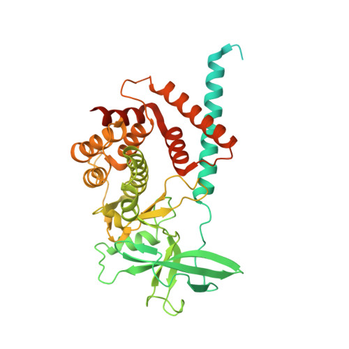
Structural insights into regulation of the PEAK3 pseudokinase scaffold by 14-3-3.

Torosyan, H.Paul, M.D.Forget, A.Lo, M.Diwanji, D.Pawlowski, K.Krogan, N.J.Jura, N.Verba, K.A.

(2023) Nat Commun 14: 3543-3543

  • DOI: https://doi.org/10.1038/s41467-023-38864-0
  • Primary Citation of Related Structures:  
    8DP5, 8DS6

  • PubMed Abstract: 

    PEAK pseudokinases are molecular scaffolds which dimerize to regulate cell migration, morphology, and proliferation, as well as cancer progression. The mechanistic role dimerization plays in PEAK scaffolding remains unclear, as there are no structures of PEAKs in complex with their interactors. Here, we report the cryo-EM structure of dimeric PEAK3 in complex with an endogenous 14-3-3 heterodimer. Our structure reveals an asymmetric binding mode between PEAK3 and 14-3-3 stabilized by one pseudokinase domain and the SHED domain of the PEAK3 dimer. The binding interface contains a canonical phosphosite-dependent primary interaction and a unique secondary interaction not observed in previous structures of 14-3-3/client complexes. Additionally, we show that PKD regulates PEAK3/14-3-3 binding, which when prevented leads to PEAK3 nuclear enrichment and distinct protein-protein interactions. Altogether, our data demonstrate that PEAK3 dimerization forms an unusual secondary interface for 14-3-3 binding, facilitating 14-3-3 regulation of PEAK3 localization and interactome diversity.


  • Organizational Affiliation

    Cardiovascular Research Institute, University of California San Francisco, San Francisco, CA, 94158, USA.


Macromolecules
Find similar proteins by:  (by identity cutoff)  |  3D Structure
Entity ID: 1
MoleculeChains Sequence LengthOrganismDetailsImage
Protein PEAK3
A, B
491Homo sapiensMutation(s): 0 
Gene Names: PEAK3C19orf35
UniProt & NIH Common Fund Data Resources
Find proteins for Q6ZS72 (Homo sapiens)
Explore Q6ZS72 
Go to UniProtKB:  Q6ZS72
GTEx:  ENSG00000188305 
Entity Groups  
Sequence Clusters30% Identity50% Identity70% Identity90% Identity95% Identity100% Identity
UniProt GroupQ6ZS72
Sequence Annotations
Expand
  • Reference Sequence
Experimental Data & Validation

Experimental Data

  • Method: ELECTRON MICROSCOPY
  • Resolution: 4.90 Å
  • Aggregation State: PARTICLE 
  • Reconstruction Method: SINGLE PARTICLE 
EM Software:
TaskSoftware PackageVersion
MODEL REFINEMENTRosetta3
RECONSTRUCTIONRELION3.1.0

Structure Validation

View Full Validation Report


Entry History & Funding Information

Deposition Data


Funding OrganizationLocationGrant Number
Other governmentQBI Independent Research Fellowship
National Institutes of Health/National Cancer Institute (NIH/NCI)United StatesU54 CA209891
National Institutes of Health/National Institute of General Medical Sciences (NIH/NIGMS)United StatesR35 GM139636

Revision History  (Full details and data files)

  • Version 1.0: 2023-06-28
    Type: Initial release