8D16

Bent ADP-Pi-F-actin

  • Classification: STRUCTURAL PROTEIN
  • Organism(s): Gallus gallus
  • Mutation(s): No 

  • Deposited: 2022-05-26 Released: 2022-09-07 
  • Deposition Author(s): Reynolds, M.J., Alushin, G.M.
  • Funding Organization(s): National Institutes of Health/National Institute of General Medical Sciences (NIH/NIGMS)

Experimental Data Snapshot

  • Method: ELECTRON MICROSCOPY
  • Resolution: 3.71 Å
  • Aggregation State: FILAMENT 
  • Reconstruction Method: SINGLE PARTICLE 

wwPDB Validation   3D Report Full Report


This is version 1.3 of the entry. See complete history


Literature

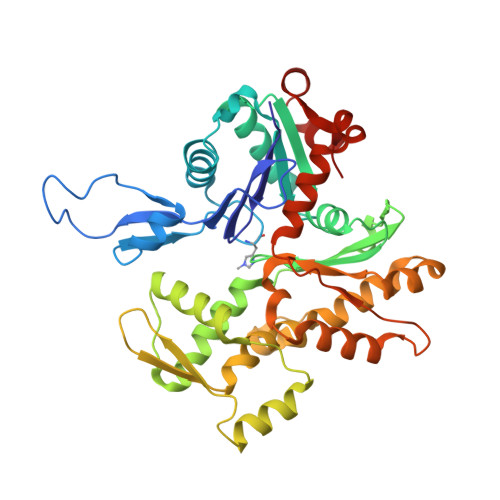
Bending forces and nucleotide state jointly regulate F-actin structure.

Reynolds, M.J.Hachicho, C.Carl, A.G.Gong, R.Alushin, G.M.

(2022) Nature 611: 380-386

  • DOI: https://doi.org/10.1038/s41586-022-05366-w
  • Primary Citation of Related Structures:  
    8D13, 8D14, 8D15, 8D16, 8D17, 8D18

  • PubMed Abstract: 

    ATP-hydrolysis-coupled actin polymerization is a fundamental mechanism of cellular force generation 1-3 . In turn, force 4,5 and actin filament (F-actin) nucleotide state 6 regulate actin dynamics by tuning F-actin's engagement of actin-binding proteins through mechanisms that are unclear. Here we show that the nucleotide state of actin modulates F-actin structural transitions evoked by bending forces. Cryo-electron microscopy structures of ADP-F-actin and ADP-P i -F-actin with sufficient resolution to visualize bound solvent reveal intersubunit interfaces bridged by water molecules that could mediate filament lattice flexibility. Despite extensive ordered solvent differences in the nucleotide cleft, these structures feature nearly identical lattices and essentially indistinguishable protein backbone conformations that are unlikely to be discriminable by actin-binding proteins. We next introduce a machine-learning-enabled pipeline for reconstructing bent filaments, enabling us to visualize both continuous structural variability and side-chain-level detail. Bent F-actin structures reveal rearrangements at intersubunit interfaces characterized by substantial alterations of helical twist and deformations in individual protomers, transitions that are distinct in ADP-F-actin and ADP-P i -F-actin. This suggests that phosphate rigidifies actin subunits to alter the bending structural landscape of F-actin. As bending forces evoke nucleotide-state dependent conformational transitions of sufficient magnitude to be detected by actin-binding proteins, we propose that actin nucleotide state can serve as a co-regulator of F-actin mechanical regulation.


  • Organizational Affiliation

    Laboratory of Structural Biophysics and Mechanobiology, The Rockefeller University, New York, NY, USA.


Macromolecules
Find similar proteins by:  (by identity cutoff)  |  3D Structure
Entity ID: 1
MoleculeChains Sequence LengthOrganismDetailsImage
Actin, alpha skeletal muscle, intermediate form
A, B, C, D, E
A, B, C, D, E, F, G
371Gallus gallusMutation(s): 0 
UniProt
Find proteins for P68139 (Gallus gallus)
Explore P68139 
Go to UniProtKB:  P68139
Entity Groups  
Sequence Clusters30% Identity50% Identity70% Identity90% Identity95% Identity100% Identity
UniProt GroupP68139
Sequence Annotations
Expand
  • Reference Sequence
Small Molecules
Ligands 3 Unique
IDChains Name / Formula / InChI Key2D Diagram3D Interactions
ADP
Query on ADP

Download Ideal Coordinates CCD File 
H [auth A]
K [auth B]
N [auth C]
Q [auth D]
T [auth E]
H [auth A],
K [auth B],
N [auth C],
Q [auth D],
T [auth E],
W [auth F],
Z [auth G]
ADENOSINE-5'-DIPHOSPHATE
C10 H15 N5 O10 P2
XTWYTFMLZFPYCI-KQYNXXCUSA-N
PO4
Query on PO4

Download Ideal Coordinates CCD File 
BA [auth G]
J [auth A]
M [auth B]
P [auth C]
S [auth D]
BA [auth G],
J [auth A],
M [auth B],
P [auth C],
S [auth D],
V [auth E],
Y [auth F]
PHOSPHATE ION
O4 P
NBIIXXVUZAFLBC-UHFFFAOYSA-K
MG
Query on MG

Download Ideal Coordinates CCD File 
AA [auth G]
I [auth A]
L [auth B]
O [auth C]
R [auth D]
AA [auth G],
I [auth A],
L [auth B],
O [auth C],
R [auth D],
U [auth E],
X [auth F]
MAGNESIUM ION
Mg
JLVVSXFLKOJNIY-UHFFFAOYSA-N
Modified Residues  1 Unique
IDChains TypeFormula2D DiagramParent
HIC
Query on HIC
A, B, C, D, E
A, B, C, D, E, F, G
L-PEPTIDE LINKINGC7 H11 N3 O2HIS
Experimental Data & Validation

Experimental Data

  • Method: ELECTRON MICROSCOPY
  • Resolution: 3.71 Å
  • Aggregation State: FILAMENT 
  • Reconstruction Method: SINGLE PARTICLE 
EM Software:
TaskSoftware PackageVersion
RECONSTRUCTIONRELION3.1

Structure Validation

View Full Validation Report


Entry History & Funding Information

Deposition Data


Funding OrganizationLocationGrant Number
National Institutes of Health/National Institute of General Medical Sciences (NIH/NIGMS)United StatesGM141044

Revision History  (Full details and data files)

  • Version 1.0: 2022-09-07
    Type: Initial release
  • Version 1.1: 2022-10-26
    Changes: Database references
  • Version 1.2: 2022-11-09
    Changes: Database references
  • Version 1.3: 2022-11-23
    Changes: Database references