8CVN

Cryo-EM Structure of Human 15-PGDH in Complex with Small Molecule SW209415


Experimental Data Snapshot

  • Method: ELECTRON MICROSCOPY
  • Resolution: 2.40 Å
  • Resolution: 2.40 Å
  • Aggregation State: PARTICLE 
  • Reconstruction Method: SINGLE PARTICLE 

wwPDB Validation   3D Report Full Report


This is version 1.1 of the entry. See complete history


Literature

Small molecule inhibitors of 15-PGDH exploit a physiologic induced-fit closing system.

Huang, W.Li, H.Kiselar, J.Fink, S.P.Regmi, S.Day, A.Yuan, Y.Chance, M.Ready, J.M.Markowitz, S.D.Taylor, D.J.

(2023) Nat Commun 14: 784-784

  • DOI: https://doi.org/10.1038/s41467-023-36463-7
  • Primary Citation of Related Structures:  
    8CVN, 8CWL, 8FD8

  • PubMed Abstract: 

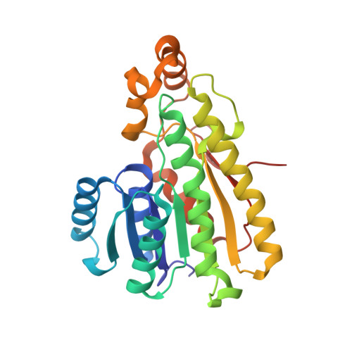
    15-prostaglandin dehydrogenase (15-PGDH) is a negative regulator of tissue stem cells that acts via enzymatic activity of oxidizing and degrading PGE2, and related eicosanoids, that support stem cells during tissue repair. Indeed, inhibiting 15-PGDH markedly accelerates tissue repair in multiple organs. Here we have used cryo-electron microscopy to solve the solution structure of native 15-PGDH and of 15-PGDH individually complexed with two distinct chemical inhibitors. These structures identify key 15-PGDH residues that mediate binding to both classes of inhibitors. Moreover, we identify a dynamic 15-PGDH lid domain that closes around the inhibitors, and that is likely fundamental to the physiologic 15-PGDH enzymatic mechanism. We furthermore identify two key residues, F185 and Y217, that act as hinges to regulate lid closing, and which both inhibitors exploit to capture the lid in the closed conformation, thus explaining their sub-nanomolar binding affinities. These findings provide the basis for further development of 15-PGDH targeted drugs as therapeutics for regenerative medicine.


  • Organizational Affiliation

    Department of Pharmacology, Case Western Reserve University, Cleveland, OH, 44106, USA.


Macromolecules
Find similar proteins by:  (by identity cutoff)  |  3D Structure
Entity ID: 1
MoleculeChains Sequence LengthOrganismDetailsImage
15-hydroxyprostaglandin dehydrogenase [NAD(+)]
A, B
256Homo sapiensMutation(s): 0 
Gene Names: HPGDPGDH1SDR36C1
EC: 1.1.1.141 (PDB Primary Data), 1.1.1 (PDB Primary Data), 1.1.1.232 (PDB Primary Data)
UniProt & NIH Common Fund Data Resources
Find proteins for P15428 (Homo sapiens)
Explore P15428 
Go to UniProtKB:  P15428
PHAROS:  P15428
GTEx:  ENSG00000164120 
Entity Groups  
Sequence Clusters30% Identity50% Identity70% Identity90% Identity95% Identity100% Identity
UniProt GroupP15428
Sequence Annotations
Expand
  • Reference Sequence
Small Molecules
Ligands 2 Unique
IDChains Name / Formula / InChI Key2D Diagram3D Interactions
NAI
Query on NAI

Download Ideal Coordinates CCD File 
C [auth A],
E [auth B]
1,4-DIHYDRONICOTINAMIDE ADENINE DINUCLEOTIDE
C21 H29 N7 O14 P2
BOPGDPNILDQYTO-NNYOXOHSSA-N
SWL (Subject of Investigation/LOI)
Query on SWL

Download Ideal Coordinates CCD File 
D [auth A],
F [auth B]
2-[butyl(oxidanyl)-$l^{3}-sulfanyl]-4-(2,3-dimethylimidazol-4-yl)-6-(1,3-thiazol-2-yl)thieno[2,3-b]pyridin-3-amine
C19 H23 N5 O S3
UNHFBUMWCMKPAQ-UHFFFAOYSA-N
Experimental Data & Validation

Experimental Data

  • Method: ELECTRON MICROSCOPY
  • Resolution: 2.40 Å
  • Resolution: 2.40 Å
  • Aggregation State: PARTICLE 
  • Reconstruction Method: SINGLE PARTICLE 

Structure Validation

View Full Validation Report


Entry History & Funding Information

Deposition Data


Funding OrganizationLocationGrant Number
National Institutes of Health/National Institute of General Medical Sciences (NIH/NIGMS)United States1RM1GM142002

Revision History  (Full details and data files)

  • Version 1.0: 2023-03-01
    Type: Initial release
  • Version 1.1: 2023-03-29
    Changes: Database references